Download
1 / 91
940 likes | 1.11k Vues
NERVOUS SYSTEM & SENSE ORGANS. Mrs. Ofelia Solano Saludar Department of Natural Sciences University of St. La Salle Bacolod City.

E N D
NERVOUS SYSTEM & SENSE ORGANS Mrs. Ofelia Solano Saludar Department of Natural Sciences University of St. La Salle Bacolod City
ORIGIN: ectoderm gives rise to the nervous system and nervous parts of sense organs • FUNCTIONS: • Regulates behavior by integrating incoming sensory information with stored information & translating that into action by way of effectors • Receives stimuli from receptors & transmits information to effectors that respond to stimulation
COMPONENTS: Billions of neurons, each of which establishes thousands of contacts with other nerve cells Neurogliacells that support, nourish, & insulate neurons Sensory receptors- integrates & stores information as memory Motor receptors- response effectors
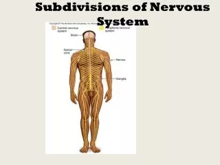
CLASSIFICATION: • Central Nervous System - brain & spinal cord • Peripheral Nervous System - cranial nerves, spinal nerves, & all their branches • Autonomic Nervous System- system of ganglia, cords & plexi controlling involuntary functions: • Sympathetic- for fight or flight response • Parasympathetic- for rest and relaxation response
ORGANIZATION (arranged dorsal to ventral in spinal cord & medulla): • Somatic sensory- sensory impulses from skin & other layers of body wall • Visceral sensory- sensations from the viscera • Visceral motor- motor impulses from smooth & branchial muscles (ANS) • Somatic motor- impulses to voluntary muscles
NERVE CELL or NEURON • Parts: • Cell body- ganglion, nucleus • Cytoplasmic processes: • Axon- also called nerve fiber, fiber tracts in CNS, nerve in PNS (sensory, motor, or mixed nerve cell processes) • Dendrite • Types: Multipolar, Bipolar, Unipolar
Parkinson’s Disease in humans is known to be caused by a decrease in dopamine (neurotransmitter) in the brain. Why not just inject patients with IV dopamine?
NEUROGLIA: supportive cells to neurons Ependymal cells – line the neurocoel and provide nutrients Oligodendroglia – produce myelin in CNS which is white and speeds up nerve impulse transmission Astrocytes – maintain blood brain barrier
Microglia – function as phagocytes • Schwann Cells – produce myelin in PNS
DEVELOPMENT OF NERVOUS SYSTEM • Neurulation • Neural tube layers: • Germinal layer – medial; with mitosis • Mantle layer – gray; cell bodies of neurons • Marginal layer – white; cytoplasmic processes without nuclei • Neuroblasts form neurons • Spongioblasts form neuroglia • Alar plate is dorsal gray matter (nuclei) • Basal plate is ventral gray matter
SUBDIVISIONS OF EMBRYONIC BRAIN • Prosencephalon (forebrain) - divides into the telencephalon & diencephalon
Mesencephalon(midbrain)- forms the tectum which includes the optic lobes (receive fibers from retina) & auditory lobes (receive fibers from inner ear) • Rhombencephalon (hindbrain) - subdivides into the metencephalon and myelencephalon
All vertebrates have a cerebrum based on the same basic plan • Major phylogenetic changes are due to loss, fusion, or enlargement of the various regions. • Phylogenetictrend in vertebrate brains is for enlargement of forebrain due to: • increasingly complex behaviors & muscle control: • coordination of limb movements more complicated (e.g., bipedal dinosaurs & birds) • increased input of sensory information & increased output of motor responses
TELENCEPHALON • Differentiates into the olfactory bulbs, tracts, lobes & cerebral hemispheres; olfactory part later becomes subordinated by cerebral hemispheres • Regions of Cerebrum: • PALLIUMhas 3 divisions: • medial (receives olfactory information) • dorsal & lateral divisions (receive other sensory input including information relayed from the thalamus)
SUBPALLIUM- consists of: • Septum - important part of the limbic system (regulates emotions & plays vital role in short-term memory) • Striatum - also called basal ganglia; present in all vertebrates & controls sequence of actions in complex movements
Agnathans, fish, & amphibians - pallia are similar • Reptiles- pallium also has a large dorsal ventricular ridge (DVR), derived from lateral pallium; DVR may be higher association area • Birds- DVR expands further; dorsal part increases in size & is called the wulst; as in reptiles, the DVR appears to serve as a higher association area • Mammals- do not have enlarged DVR but the dorsal pallium is enlarged & is called the CEREBRAL CORTEX; cortex receives & analyzes sensory information & initiates motor activity
CEREBRAL STRIATUM COMPLEX: • Paleostriatum– primary region in fish, primarily involved with olfactory reflexes • Neostriatum – beginning with reptiles, more complex and paleostriatum becomes buried • Hyperstriatum – primarily in birds responsible for stereotypical behavior (migration, courting, nesting) • Corpus striatum or basal nuclei – paleostriatum buried in mammalian brain, responsible for stereotyped & repetitive movements • Primitive pallium displaced by expansion of NEOCORTEX
CEREBRAL CORTEX • Becomes increasingly folded to fit in skull starting with reptiles up to mammals • Functions: voluntary movement (motor), conscious sensations (sensory), memory, integration (decisions) • 4 lobes according to skull bones
DIENCEPHALON- differentiates into: • Hypothalamus– optic chiasma, infundubular stalk for pituitary gland & mammmilary bodies particularly well developed in fishes • Ventral part of infundibulum unites with Rathke’s pouch to form the pituitary • In fishes, infundibulum also includes the inferior lobes & vascular sac • functions as endocrine organ, regulates ANS, sexual & emotional behavior, water balance, thermostat, hunger, satiety • Thalamus – relay center for sensory impulses from all parts of the body
Epithalamus- dorsal part of diencephalon; gives rise to the pineal body (epiphyses) which functions as: • A light receptor in agnathans & affects skin pigmentation in lower vertebrates • An endocrine organ in gnathostomes; plays a role in regulating biological rhythms
MESENCEPHALONcomposed of: • Optic lobes – 2 in most vertebrates, 4 in mammals, especially well developed in birds • Auditory lobes – auditory reflexes • Corpora Quadrigemina • Cerebral Peduncles – motor tracts • Cerebral Aqueduct – for CSF
METENCEPHALON • Small in cyclostomes & amphibians but increases in size & importance in amniotes • Consists of: • Pons - pathway for ascending & descending fiber tracts & origin of cranial nerves V, VI, & VII • Cerebellum - modifies & monitors motor output; important in maintaining equilibrium
MYELENCEPHALON • Undergoes little change in the vertebrate series • Connection between brain & spinal cord for ascending & descending pathways • Consists of the medulla oblongata & its major functions include: • origin of cranial nerves (VII - X or VII - XII) • contains centers important in regulating respiration, heartbeat, & intestinal motility
VENTRICLES- cavities containing cerebrospinal fluid (CSF) providing cushion, protection, nutrients • Formed from blood vessels called choroid plexus • Telachoroidea- covers the ventricles & from which choroid plexi project into ventricles • Location: 1st & 2nd lateral ventricles- cerebrum; 3rd ventricle- diencephalon; 4th ventricle- medulla
CRANIAL NERVES • Most fishes, amphibians have 10 cranial nerves; crossopterygians & amniotes have 12 • Sensory fibers have their cells of origin in external ganglia on the nerve roots. • Sensory & motor roots do not unite; cranial nerves are irregular as to their functional components.