Download
1 / 33
440 likes | 1.24k Vues
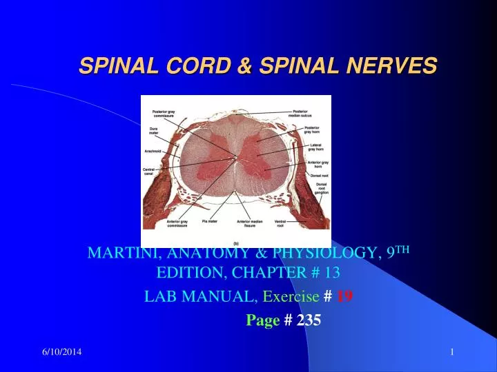
SPINAL CORD & SPINAL NERVES. MARTINI, ANATOMY & PHYSIOLOGY, 9 TH EDITION, CHAPTER # 13 LAB MANUAL, Exercise # 19 Page # 235. NOTE:. THIS IS A STUDY GUIDE , NOT AN ALL INCLUSIVE REVIEW.

E N D
SPINAL CORD & SPINAL NERVES MARTINI, ANATOMY & PHYSIOLOGY, 9TH EDITION, CHAPTER # 13 LAB MANUAL, Exercise#19 Page# 235
NOTE: • THIS IS A STUDY GUIDE, NOT AN ALL INCLUSIVE REVIEW. • THERE MIGHT BE THINGS NOT COVERED BY THIS STUDY GUIDE THAT MIGHT BEASKED IN YOUR PRACTICUMS / QUIZZES. • STUDENTS ARE RESPONSIBLE FOR READING THEIR TEXBOOK (S) AND FOR ALL THE MATERIAL COVERED DURING THE LABORATORY PERIOD, AS PER THE COURSE SYLLABUS
Competency 6: The Nervous System Upon successful completion of this laboratory, the students will be able to demonstrate an understanding of the structural and functional features of the nervous system including the special sense organs by: Describing the structure and functions of the spinal cord and the spinal nerves and their plexuses.
SPINAL MENINGES • Surrounds the spinal cord for physical stability • & Shock absorption • Wall of vertebral canal (no part of meninges) • Epidural space- contains fat and blood vessels • imp for anesthetic injection • Dura mater- outermost covering of spinal cord • Subdural space • Aracnoid mater • Subarachnoid space- filled with CSF • CSF- shock absorber • diffusion medium for wastes, gases, • nutrients & chemicals • Pia mater-pia=delicate, mater=mother for nourishment
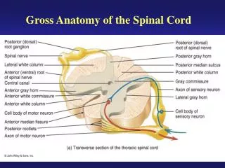
SPINAL CORD • Dendicular ligaments- prevent lateral movement of the spinal cord • Conus medularis- • End part of the spinal cord, like a conus • Cauda equina- nerve fibers extending after • conus medularis, like a horse tail • Filum terminale- to anchor spinal cord vertically by connecting it with coccyx • F- to preventup & down movement • Gray matter- it has nuclei • Posterior (dorsal) horns –somatic & visceral sensory nuclei • Anterior (ventral) horns-somatic motor nuclei • Lateral horns- autonomic (visceral) nuclei
Grey commissure- to connect two sides of the grey mater • Central canal (the hole in the middle)- • Conteins CSF(cephalo spinal fluid) • White matter- it has tracs or fasciculus • Posterior (dorsal) white column (funiculus) • Carry sensory info • Lateral white column (funiculus)- • Carry sensory & motor info • Anterior (ventral) white column (funiculus) • Carry sensory & motor info • Anterior median fissure- deep groove • Posterior median sulcus- shallow groove
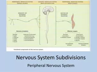
SPINAL NERVES • Posterior root- carry sensory info • Posterior root ganglia- conteins sensoryneurons • Anterior root- carries motor info • Posterior ramus (smaller)- carries sensory & motorinfo • Anterior ramus- (thicker)- carries sensory & motorinfo
PLEXUSES • CERVICAL • BRACHIAL • LUMBAR • SACRAL
Peripheral Nerves and Plexuses ALFONSO A. PINO. MD.
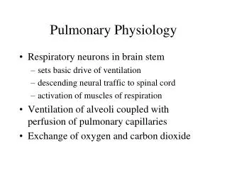
CERVICAL PLEXUS • Serves or innervates neck & shoulder • Phrenic nerve- it extends below the shoulder to the • diaphragm. • It causes the diaphragm to contract • important for breathing
Brachial plexus- serves or innervates upper limb and shoulder • Axillary n- serves deltoid & teres minor • And skin of the shoulder • (From anterior to posterior)- • Radial nerve- serves posterior upper limb • Musculocutaneous n-serves anterior arm, • biceps brachii & brachialis • Median n- serves anterior forearm • Ulnar n- serves anterior hand
Spinal Nerves and Plexuses The Brachial Plexus of the Ventral Rami Major nerves of brachial plexus Musculocutaneous nerve(lateral cord) Median nerve (lateral and medial cords) Ulnar nerve (medial cord) Axillary nerve (posterior cord) Radial nerve (posterior cord)
LUMBAR PLEXUS • It serves or innervates skin & some muscles of anterior abdomen, buttocks, genitalia, anterior lateral & posteromedial thigh & medial surface of leg and foot • Femoral nerve: anterior muscles of the thigh, flexors & adductors of the hip, skin of the thigh(antero medial surface) & skin over the medial surface of leg and foot • RECTUS FEMORIS • VASTUS MEDIALIS • VASTUS LATERALIS • VASTUS INTERMEDIUS
LUMBAR PLEXUS & SACRAL PLEXUS ALFONSO A. PINO. MD.
SACRAL PLEXUS • It serves or innervates: buttock (gluteus) & • posterior lower limb, anterior leg & foot • lateral leg, some muscles of genitalia • including external anal and urethral sphincters • Sciatic n (thiker)- serves posterior thigh • Common peroneal n (goes anteriorly)- serves anterior leg & foot • Tibial n (goes posteriorly)- serves posterior leg and foot
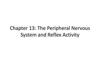
Components of a Reflex Arch ALFONSO A. PINO. MD.
The Patellar Reflex ALFONSO A. PINO. MD.
REMEMBER!GO TO THE TUTORING ROOMAND PRACTICE WITH MODELS.ROOM 3326.