Download
1 / 29
290 likes | 296 Vues
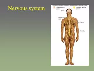
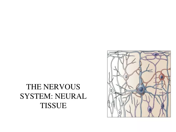
THE NERVOUS SYSTEM: NEURAL TISSUE. Cells in Nervous Tissue. Neurons Neuroglia. Neuroglia (Glia). about half the volume of cells in the CNS smaller than neurons 5 to 50 times more numerous do NOT generate electrical impulses divide by mitosis 2 types in PNS Schwann cells

E N D
Cells in Nervous Tissue • Neurons • Neuroglia
Neuroglia (Glia) • about half the volume of cells in the CNS • smaller than neurons • 5 to 50 times more numerous • do NOT generate electrical impulses • divide by mitosis • 2 types in PNS • Schwann cells • Satellite cells • 4 types in the CNS • Astrocytes • Oligodendrocytes • Microglia • Ependymal cells
Astrocytes • Largest of glial cells • Star shaped with many processes projecting from the cell body • Help form and maintain blood-brain barrier • Provide structural support for neurons • Regulate the chemical/ion environment for generation of nerve impulse • Regulate nutrient & ion concentrations for neuron survival • Take up excess neurotransmitters
Oligodendrocytes • Most common glial cell type • Each forms myelin sheath around the axons of neurons in CNS • Analogous to Schwann cells of PNS • Form a supportive network around CNS neurons
Microglia • few processes • derived from mesodermal cells that also give rise to monocytes and macrophages • Small cells found near blood vessels • Phagocytic role - clear away dead cells • protect CNS from disease through phagocytosis of microbes • migrate to areas of injury where they clear away debris of injured cells - may also kill healthy cells
Ependymal Cells • epithelial cells arranged in a single layer • range in shape from cuboidal to columnar • line ventricles of the brain & central canal of spinal cord • produce & circulate the cerebrospinal fluid (CSF) • CSF = colorless liquid that protects the brain and SC against chemical & physical injuries, carries oxygen, glucose and other necessary chemicals from the blood to neurons and neuroglia
PNS: Satellite Cells • Flat cells surrounding PNS neuronal bodies • hold the cell bodies together to form a ganglion
PNS: Schwann Cells • produces part of the myelin sheath surrounding an axon in the PNS • contributes regeneration of PNS axons
Neurons • have the property of electrical excitability - ability to produce action potentials or nerve impulses in response to stimuli
Representative Neuron http://www.horton.ednet.ns.ca/staff/selig/Activities/nervous/na1.htm 1. cell body or soma -same components of a typical eukaryotic cell -e.g. nucleus, Golgi, mitochondria -Nissl bodies -rough ER & ribosomes for protein synthesis -cytoskeleton of neurofilaments and microtubules to give neuron it’s shape and to move neurotransmitters to the terminals
Neurons 2. Cell processes = dendrites (little trees) - the receiving or input portion of the neuron -short, tapering and highly branched -surfaces specialized for contact with other neurons
3. Cell processes = axon • conducts nerve impulses away from cell body to another neuron • joins the cell body at a cone-shaped elevation = axon hillock • nerve impulse arises at a region of the axon hillock = trigger zone • cytoplasm = axoplasm • plasma membrane = axolemma • side branches = collaterals arise from the axon • axon and collaterals end in fine processes called axon terminals • swollen tips called synaptic end bulbs contain vesicles filled with neurotransmitters
Classification of Neurons • neurons can be classified based on: • their shape – e.g. multipolar, bipolar, unipolar • who identified them – e.g. Purkinje • function • Sensory (afferent) neurons • transport sensory information from skin, muscles, joints, sense organs & viscera to CNS • Motor (efferent) neurons • send motor nerve impulses to muscles & glands • Interneurons (association) neurons • connect sensory to motor neurons • 90% of neurons in the CNS
The Nerve Impulse: Terms to know • membrane potential = electrical voltage difference measured across the membrane of a cell • results from the build-up of negative ions in the cytosol along the inside of the neuron’s PM • the outside of the PM becomes more positive • this difference in charge can be measured as potential energy – measured in millivolts • resting membrane potential = membrane potential of a neuron measured when it is unstimulated • ranges from -40 to -90 mV
The Nerve Impulse: Terms to know • polarization – change in membrane potential • 1. depolarization – increase in MP away from resting • 2. hyperpolarization – decrease in MP away from resting • 3. repolarization – “return to resting membrane potential”
Ion Channels • ion channels in the PM of neurons and muscles contributes to their excitability • when open - ions diffuse down their concentration gradients • some ion channels are permanently open – non-gated channels • some ion channels possess gates to open and close them – gated channels • two types: ligand gated & voltage gated
Ion Channels 1. Leakage (non-gated) or Resting channels: are always open, contribute to the resting potential -nerve cells have more K+ than Na+ leakage channels -so K+ leak channels contribute more to resting membrane potential than Na+ leak channels -leaking ions are pumped back to where they belong 2. Gated channels: open and close in response to a stimulus A. voltage-gated: open in response to change in voltage - participate in the AP B. ligand-gated: open & close in response to particular chemical stimuli (hormone, neurotransmitter, ion) C. mechanically-gated: open with mechanical stimulation
Action Potential • Resting membrane potential is -70mV • AP triggered when the membrane potential reaches a threshold usually -55 MV • if the membrane potential exceeds that of threshold Action Potential • action potential = nerve impulse • takes place in two stages: depolarizing phase (more positive) and repolarizing phase (more negative - back toward resting potential) • followed by a hyperpolarizing phase or refractory period in which no new AP can be generated http://www.blackwellpublishing.com/matthews/channel.html
Action Potential 6. • 1. neuron is at resting membrane potential (resting MP) • 2. neuron binds neurotransmitters via ligand-gated sodium channels • 3. channels open & Na diffuses into neuron = depolarization • inside of neuron (i.e. MP) becomes more positive • 4. if neuron depolarizes enough & reaches Threshold Action Potential (AP) • 5. 1st stage of AP – opening of voltage-gated Na channels • large diffusion of Na+ ions into neuron = BIG depolarization • membrane potential goes from negative to positive 4. 5. 7. 1. 3. • 6. closing of VGNa channels & opening of voltage-gated K channels • 7. BIG outflow of potassium through VGK channels = repolarization • inside of neuron (MP) becomes more negative • 8. closing of VGK channels BUT so much K+ has diffused out – neuron’s MP goes past resting and hyperpolarizes • 9. neuron is hyperpolarized – no new AP can be generated with a normal stimulus • 10. all voltage-gated channels closed, Na/K pump “resets” ion distribution to resting situation 8. 10. 9. 2.
Continuous versus Saltatory Conduction • Continuous conduction (unmyelinated fibers) • action potential spreads continuously over the surface of the axolemma • as one section of the axon is depolarized, the membrane potential of the next section is depolarized toward threshold http://highered.mcgraw-hill.com/sites/0072437316/student_view0/chapter45/animations.html#
Saltatory Conduction • Saltatory conduction -depolarization happens only at Nodes of Ranvier - areas along the axon that are unmyelinated and where there is a high density of voltage-gated ion channels -action potential “jumps” from node to node http://www.blackwellpublishing.com/matthews/actionp.html
Synapses • Synapse: Site of intercellular communication between 2 neurons or between a neuron and an effector (e.g. muscle – neuromuscular junction) • Permits communication between neurons and other cells • Initiating neuron = presynaptic neuron • Receiving neuron = postsynaptic neuron • You can classify a synapse according to: • 1. the action they produce on the post-synaptic neuron – excitatory or inhibitory • 2. the mode of communication – chemical vs. electrical
Synapses • Electrical • Direct physical contact between cells required • Conducted through gap junctions • Two advantages over chemical synapses • 1. faster communication – almost instantaneous • 2. synchronization between neurons or muscle fibers • e.g. heart beat
Chemical Synapse • Most common type of synapse • Membranes of pre and postsynaptic neurons do not touch • Space = Synaptic cleft • Most are axon terminal dendrite • Some are axon terminal axon http://www.lifesci.ucsb.edu/~mcdougal/neurobehavior/modules_homework/lect3.dcr
Chemical Synapse • the AP cannot travel across the cleft – release of neurotransmitters • 1. arrival of action potential in the synaptic end bulb • 2. opening of voltage-gated calcium channels – influx of Ca2+ into end bulb • 3. docking of synaptic vesicles with NTs with plasma membrane – release of NTs into synaptic cleft • 4. binding of NT to ligand-gated channels – channels open • 5. diffusion of Na+ ions into post-synaptic membrane • 6. depolarization of post-synaptic neuron – if the NT is excitatory • 7. depolarization to threshold Action Potential • if the neurotransmitter is an inhibitory NT - then the post-synaptic neuron will hyperpolarize rather than depolarize • NO ACTION POTENTIAL!!! http://www.blackwellpublishing.com/matthews/nmj.html
The Neuromuscular Junction • the motor neuron’s synaptic terminal is in very close association with the muscle fiber • distance between the bulb and the folded sarcolemma = neuromuscular junction • neurotransmitter released = acetylcholine https://www.youtube.com/watch?v=7wM5_aUn2qs