Download
1 / 1
10 likes | 102 Vues
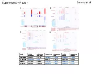
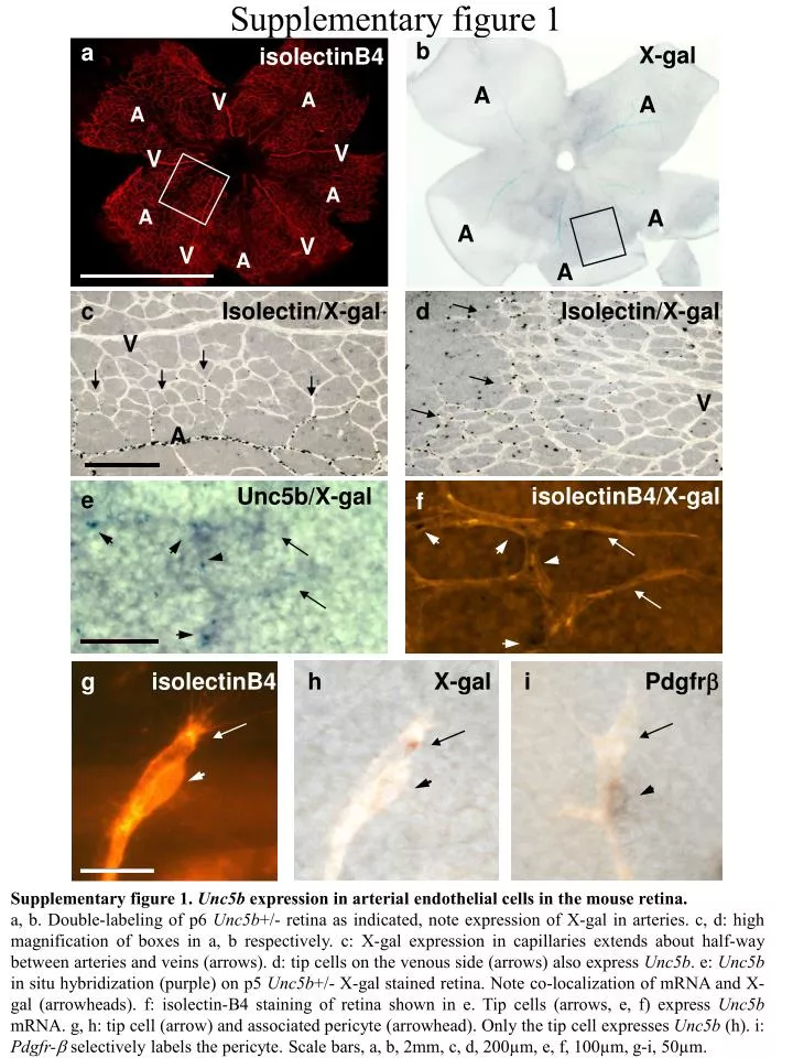
Supplementary figure 1. b. a. X-gal. isolectinB4. A. V. A. A. A. V. V. A. A. A. A. V. V. A. A. c. Isolectin/X-gal. d. Isolectin/X-gal. V. V. A. Unc5b/X-gal. isolectinB4/X-gal. e. f. g. isolectinB4. h. X-gal. i. Pdgfr b.

E N D
Supplementary figure 1 b a X-gal isolectinB4 A V A A A V V A A A A V V A A c Isolectin/X-gal d Isolectin/X-gal V V A Unc5b/X-gal isolectinB4/X-gal e f g isolectinB4 h X-gal i Pdgfrb Supplementary figure 1. Unc5b expression in arterial endothelial cells in the mouse retina. a, b. Double-labeling of p6 Unc5b+/- retina as indicated, note expression of X-gal in arteries. c, d: high magnification of boxes in a, b respectively. c: X-gal expression in capillaries extends about half-way between arteries and veins (arrows). d: tip cells on the venous side (arrows) also express Unc5b. e: Unc5b in situ hybridization (purple) on p5 Unc5b+/- X-gal stained retina. Note co-localization of mRNA and X-gal (arrowheads). f: isolectin-B4 staining of retina shown in e. Tip cells (arrows, e, f) express Unc5b mRNA. g, h: tip cell (arrow) and associated pericyte (arrowhead). Only the tip cell expresses Unc5b (h). i: Pdgfr-b selectively labels the pericyte. Scale bars, a, b, 2mm, c, d, 200µm, e, f, 100µm, g-i, 50µm.