Download
1 / 2
20 likes | 168 Vues
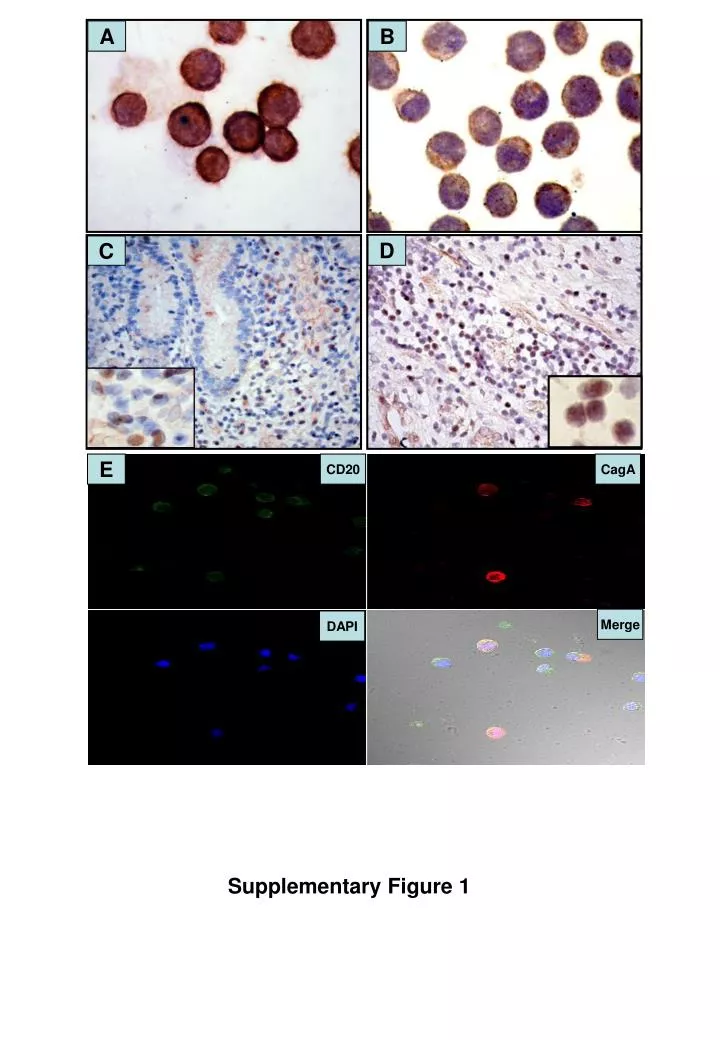
A. B. D. C. E. CD20. CagA. Merge. DAPI. Supplementary Figure 1.

E N D
A B D C E CD20 CagA Merge DAPI Supplementary Figure 1
Supplementary Figure 1. Immunohistochemical analysis of CagA and phopho-SHP-2 expression in human B cells from the CagA-translocated cell line and H. pylori-positive gastric MALT lymphoma tissues (A) Expression of CagA in cells of the CagA-translocated human B cell line. (B) Absence of CagA expression in cells of the human B cell line. (C) An H. pylori-positive gastric MALT lymphoma case displaying CagA expression in tumor cells of the gastric mucosa (right bottom inset, 1000×). (D) An H. pylori-positive gastric MALT lymphoma case displaying phospho-SHP-2 (Thy542; AF3790, R&D Systems, Minneapolis, MN) expression in tumor cells of the gastric mucosa (left bottom inset, 1000×). (E) Confocal microscopy showing that most CagA-positive B cells from the CagA-translocated human B cell line (red fluorescence)express CD20 (green fluorescence).