Download
1 / 58
580 likes | 1.17k Vues
Management of Gastrointestinal Foreign Bodies. William J. Fecht, Jr., M.D. Indiana Gastroenterology, Inc. www.indianagastro.com March 5, 2011. Course Objectives. Recognize indications for emergent, urgent and elective endoscopic management.

E N D
Management of Gastrointestinal Foreign Bodies William J. Fecht, Jr., M.D. Indiana Gastroenterology, Inc. www.indianagastro.com March 5, 2011
Course Objectives Recognize indications for emergent, urgent and elective endoscopic management. List equipment that should be readily available to facilitate foreign body removal. Summarize the types of foreign bodies and techniques to facilitate removal. If it is within your means to make a childhood dream come true, don’t hesitate … go for it!
Case 1 A 30 year old otherwise healthy male presents complaining of progressive right upper quadrant pain. CT scan is performed revealing thickening of the duodenal sweep. This leads to a small bowel follow through which identifies a long slender object in his proximal bowel. Patient recalls swallowing a toothpick 2 weeks ago while enjoying a cold one. Consent is obtained and he proceeds to upper endoscopy …
Case 1 • Pediatric colonoscope • 4th portion of duodenum • Forceps • Snare • 2 weeks oral antibiotics • Cipro / Flagyl • Diet as tolerated
Duodenal Foreign Body The DAVE Project (www.darveproject.org) Peter B. Kelsey, M.D. Harvard Medical SchoolMassachusetts General Hospital
Case 2 43 year old institutionalized male with severe mental retardation presents for open access upper endoscopy to evaluate new onset non-bilious vomiting. Upper endoscopy is performed …
Case 2 • 2 cm esophageal FB encountered at 33 cm • Overtube • Rat tooth forceps • Cap insulators (2) • Remove all potential FBs from patient’s reach
Case 3 82 year old male well known to the endoscopy staff who undergoes frequent dilations for quinidine induced esophageal stricture. He reports abrupt onset of dysphagia and inability to tolerate oral secretions while eating a turkey dinner. On exam, he is in no distress but frequently spitting into a cup. Abdomen is benign. 0.5 mg Glucagon is given and symptoms improve for approximately a half-an-hour. Consent is obtained and patient undergoes upper endoscopy …
Case 3 • FB found at 35 cm – site of known stricture • Overtube placed • Gently advanced into the stomach • 18 mm TTS balloon dilation • Tolerates liquid then regular diet • Discharged to home on same day
Case 4 While walking through the MICU, the GI attending overhears a conversation regarding a patient who ingested a foreign body with life threatening toxicity which has failed to pass despite 48 hours of observation and 2 gallons of Go-lytely. “Dag bernit,” he says under a hushed voice. This is a emergency. Call Jaime now and get the cart down here. Patient does not report any obstructive symptoms. On exam he appears comfortable and the abdomen is benign. He is hesitant to sign the consent as he contemplates possible death to felony charges but ultimately agrees. Upper endoscopy is performed …
Case 4 • Foreign body is seen floating in a pool of prep • Endoscope is removed • Surgery • FB is submitted to path for gross review • “multiple irregularly shaped hard white rocks contained within a ‘knotted’ bag” - LBM
Dr. Chevalier Jackson Laryngologist Invented the modern science of endoscopy of the upper airway and esophagus. Pioneered methods for safer removal of foreign bodies which previously had been associated with extremely high mortality. Died August 16, 1958 and obituary read “one of the greatest, if not the greatest laryngologists of all time.”
What is this object? Toy opera glasses
Patient Profiles • Pediatric population (6 months to 3 years) • Coins, keys, buttons, nails, pins, batteries • Adult population • Intentional ingestion • Psychiatric patients, secondary gain • Accidental ingestion • Elderly, Demented, Intoxicated • Esophageal pathology • Iatrogenic • Capsule endoscopy
Food Bolus Impaction • Most common GIFB that comes to medical attention in adults. • Predisposing factors • 75 to 100% will have esophageal pathology • Schatzki’s (“B”) Rings • Peptic strictures • Spasm • Webs • Extrinsic compression • Surgical: Anastomoses, Nissen wraps, gastroplasties • Esophageal cancer presenting as an acute GIFB is uncommon but recognized
Numbers • 80% to 90% of GIFBs pass spontaneously • Mortality • 50% in early 20thcentury • If the foreign body didn’t kill you the chest surgery often did. • 1,500 deaths per year (Arch Surg 1977) • Complications • Most common: Bowel perforation and obstruction • Other: Bleeding, Abscess, Fistula, Respiratory • 5% perforation rate • 35% for sharp objects
Sites of impaction • Anatomic sphincter • Acute angulation • Physical narrowing • Prior surgery • Congenital gut malformations
Esophagus • 4 areas of physiologic narrowing • UES and cricopharyngeal muscle • Aortic arch • Crossing of main stem bronchus • Gastroesophagealjunction • Esophageal pathology further enhance impaction • Motor disturbances • Achalasia, spasm • Motility?
Stomach • 80% to 90% of all ingested objects pass once in the stomach • Objects which are unlikely to pass • Size and shape • Long objects > 5 cm • Large diameter objects > 2 cm • Pyloric disease • Retrieval indicated for objects which fail to pass in 5 to 7 days
Small intestine • Long and pointed objects hang up in duodenal sweep • Ligament of Treitz • Objects trapped at ileocecal valve tend to be smaller • It is rare for foreign bodies to lodge or cause complications in the small bowel • Axial flow and peristalsis orients object favorably • Surrounded by stool
History, Exam, Imaging diagnosis
History • Careful history • Estimation of suspected level of impaction is unreliable • EXCEPTION: Cricopharyngeal muscle • Children, Noncommunicative or mentally impaired • Requires high index of suspicion • May be witnessed • Choking, refusal to eat, vomiting, drooling, wheezing, blood-stained saliva, respiratory distress • Past medical history • Previous GIFB impactions • Alcohol use • Occupational history
Physical Examination • Attention to the neck, respiratory status and abdomen • Signs of perforation require surgical intervention • Swelling, erythema, tenderness or crepitus in the neck region • Peritoneal signs (guarding w/ rebound) on abdominal examination • Airway assessment • Oxygenation • Ventilation • Aspiration risk?
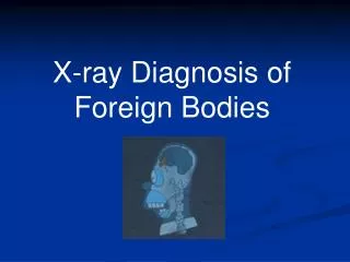
Radiographic studies • AP and Lateral Radiographs • Chest and Abdomen • Neck if hypopharynx or cervical esophagus • Contrast studies play limited role and should be avoided. • Avoid barium if perforation suspected • Role if symptoms unclear or nonspecific and endoscopy is relatively contraindicated • CT scanning may be useful in some situations • Handheld metal detectors • Persistent symptoms related to the esophagus in caes of suspected FB ingestion should be pursued with endoscopy even after unrevealing radiographic evaluation.
Nonendoscopic Therapies • Smooth muscle relaxants • Glucagon (0.5 to 2 mg) • 12% to 58% reported success • Nifedipine • Nitroglycerin • Gas-forming agents • 75% - 100% success rates • Esophageal rupture / perforation • C/I: Fixed rigid obstruction, > 6 hr, proximal 1/3 of esophagus • Historical • Papain (meat tenderizer) • Emetics • Radiographic / Fluoroscopic guidance • Balloon catheters, Wire baskets, Suction catheters • Magnet catheters
Timing of Endoscopy • Emergent • Features of high-grade esophageal obstruction or acute distress • Sharp and pointed objects • Disc battery lodged in the esophagus • Urgent • Suspected foreign body • Most food impactions • No high-grade obstruction and not in acute distress • Under no circumstances should a FB be allowed to remain in the esophagus beyond 24 hours. • Elective • Following passage or extraction when symptoms persist • Laceration, retained foreign material • Evaluate for underlying esophageal pathology • Not indicated • Small, blunt objects which have passed into stomach • Contraindicated • Bowel perforation • Small bowel obstruction beyond Ligament of Treitz
Endoscopic equipment • Endoscopes • Standard and therapeutic • Laryngoscope • Transnasally inserted bronchoscope • Overtubes • Airway protection • Potential for multiple passes • Protection of esophagus from objects • Less commonly used in children due to risk of trauma • Accessory Equipment • Polypectomy snare • Dormia basket • Roth retrieval net • Foreign body rat tooth or alligator forceps • Stiegmann-Goff friction fit variceal adapter • Latex protector hood
Specific Management of GIFBs • Food bolus impaction • Coins and other small blunt objects • Sharp/Pointed and Long objects • Narcotic packets
Food Bolus Impaction • Most common GIFB in adults • Urgent management when severe distress, excessively salivating or unable to manage secretions • Alleviate within 12 to 24 hours of occurrence • Management . . .
Management • Glucagon 1 mg IV • Many pass with gentle nudge of endoscope • Forceful blinded pushing is never indicated • Disrupt and debulk with forceps then advance • En toto or piecemeal removal with grasping forceps • Variceal banding device • Hot dogs, chicken meat, difficulty grasping food • Unsuccessful • Repeated attempt by second endoscopist • Rigid esophagoscopy • Laparotomy / Thoracotomy
Coins and other blunt objects • Dimes and pennies – usually pass • Prompt removal if esophageal impaction • Pressure necrosis, perforation, fistulization otherwise • Management • Esophageal impaction • Grasping forceps • Advance into stomach and retrieve with Roth net or Dormia basket • Stomach • Conservative outpatient management if < 25 mm • Regular diet and observe stools • Weekly radiographs • Endoscopic removal if persists up to 4 weeks • Post pyloric and fail to progress or symptomatic • Surgical management
Long Objects Longer than 6 to 10 cm will have difficulty passing the duodenal sweep. Consider use of a longer (>45 cm) overtube. Use snare or basket and maneuver object into overtube.
Sharp / Pointed Objects • Most dangerous and most challenging • Account for 1/3 of all GIFB perforations • Esophageal impaction is emergent • Animal bones and toothpicks are most common GIFBs requiring surgery in U.S. • Management • Direct laryngoscopy for objects lodged at or above cricopharyngeus. • Polypectomysnares and FB retrieval forceps • “Advancing points puncture, trailing points do not” • Latex protector hood • Surgery if object fails to pass in 3 days or symptomatic
Disk Batteries • Liquefaction necrosis and perforation can occur rapidly with esophageal impactions. • Radiographs to localize esophagus vs. stomach • Endoscopic emergency for esophageal impactions. • Batteries in the stomach can be observed unless signs / symptoms or injury, over 20 mm in size or fail to pass after 48 hours. • Management • Stone retrieval basket or roth net • If battery cannot be retrieved from esophagus it should be advanced into stomach. • Emetics and acid suppressive therapy have no role. • GI lavage may expedite passage.
Narcotic Packets • “Body stuffer” vs. “Body packer” or “mule” • Cocaine and heroin • Abdominal radiographs or CT • Toxicology screens may suggest leakage • Endoscopic removal is absolutelycontraindicated • Iatrogenic rupture of leakage of contents is fatal. • Management • Observation (inpatient) with clear liquid diet • Asymptomatic • GI lavage? • Surgery • Obstruction, fails to progress or suspected rupture
St. Louis University Experience Luxon-Fecht 9/02 Prather-Fishman 6/01 Tetri-Fazel 11/98 Befeler-Pappas 5/02