Download
1 / 11
120 likes | 294 Vues
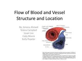
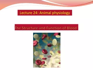
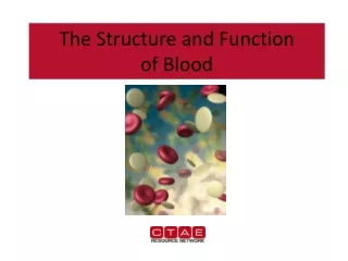
Flow of Blood and Vessel Structure and Location . Ashley Meyer, Ashley Baker, King, Quentin Key . Major Veins and Arteries . Aorta Left Pulmonary Artery Right Pulmonary Artery Pulmonary veins Superior Vena Cava Inferior Vena Cava Coronary Arteries Right Atrium Right Ventricle

E N D
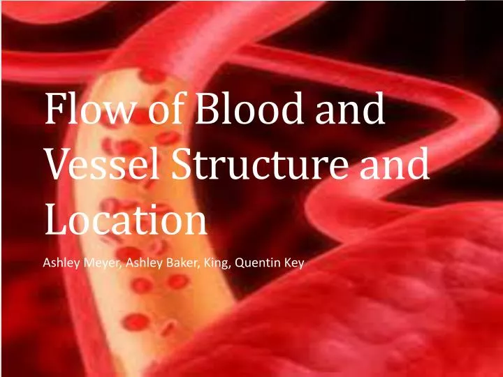
Flow of Blood and Vessel Structure and Location Ashley Meyer, Ashley Baker, King, Quentin Key
Major Veins and Arteries • Aorta • Left Pulmonary Artery • Right Pulmonary Artery • Pulmonary veins • Superior Vena Cava • Inferior Vena Cava • Coronary Arteries • Right Atrium • Right Ventricle • Left Atrium • Left Ventricle
Major Arteries • Left Coronary Artery –Divides into two branches: the circumflex artery and the left anterior descending artery • Circumflex Artery– Supplies blood to the left atrium and the side and back of the left ventricle • Left Anterior Descending Artery (LAD) –The "LAD", or left anterior descending artery (or anterior interventricular branch of the left coronary artery, or anterior descending branch) is an artery of the heart that supplies blood to the front and bottom of the left ventricle and the front of the septum • Right Coronary Artery (RCA) –Supplies blood to the Right Atrium, right ventricle, and bottom of the septum • Coronary Veins – takes oxygen-poor or deoxygenated blood that has already been “used” by muscles of the Heart and return it to the right atrium • Vena Cava – The superior and inferior vena cava are collectively called the venae cavae. They are the veins that return deoxygenated blood from the body into the heart. They both empty into the right atrium • Aorta– The largest artery in the body, originating from the left ventricle of the heart and extending down to the abdomen, where it branches off into two smaller arteries (the common iliacs). The aorta distributes oxygenated blood to all parts of the body through the systemic circulation • Pulmonary Artery – The pulmonary arteries carry blood from the heart to the lungs. They are the only arteries (other than umbilical arteries in the fetus) that carry deoxygenated blood
Major Veins • Small Cardiac Vein- An inconstant vessel that accompanies the right coronary artery in the coronary sulcus from the right ventricle and empties into the coronary sinus or the middle cardiac vein. • Oblique Vein - A tributary of the coronary sinus; on the posterior wall of the left atrium • Coronary Sinus – A short trunk receiving most of the cardiac veins, beginning at the junction of the great cardiac vein and the oblique vein of the left atrium, running in the posterior part of the coronary sulcus and emptying into the right atrium between the inferior vena cava and the atrioventricular orifice. • Great Cardiac Vein – AKA the left coronary vein. A tributary of the coronary sinus that begins at the apex of the heart and runs in the anterior interventricular sulcus. • Veins from the Left Ventricle – • Middle Cardiac Vein –
Misc. Vocab • Atrium – A body cavity or chamber, especially either of the upper chambers of the heart that receives blood from the veins and forces it into a ventricle. Also called auricle • Coronary – Of or relating to the heart • Apex – The highest point; the vertex • Sulcus – A groove, trench, or furrow; in anatomy, a general term for such a depression, especially one on the brain surface, separating the Gyri • Gyri – Plural of Gyrus, rounded ridge, as on the surfaces of the cerebral hemispheres • Cerebral – Pertaining to the cerebrum • Cerebrum – The main portion of the brain, occupying the upper part of the cranial cavity; its two hemispheres, united by the corpus callosum, form the largest part of the central nervous system in humans. The term is sometimes applied to the postembryonic forebrain and midbrain together or to the entire brain • Atrioventricular –Pertaining to both an atrium and a ventricle of the heart • Ventricle – A small cavity or chamber, as in the brain or heart • Corpus Callosum– The commissural plate of nerve fibers connecting the two cerebral hemispheres except for most of the temporal lobes. Also called commissure of cerebral hemispheres • Commissural – Relating to a commissure • Commissure –A site of union of corresponding parts; specifically, the sites of junction between adjacent cusps of the heart valves • Postembryonic – Following the embryonic stage of development • Cranial – Of or relating to the skull or cranium • Cusp – A triangular fold or flap of a heart valve • Aortic – The main trunk of the systemic arteries, carrying blood from the left side of the heart to the arteries of all limbs and organs except the lungs
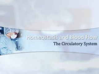
Flow of blood through the heart • Blood from the body flows: • to the superior and inferior vena cava, • then to the right atrium • through the tricuspid valve • to the right ventricle • through the pulmonic valve • to the pulmonary artery • to the lungs • The blood picks up oxygen in the lungs, and then flows from the lungs: • to the pulmonary veins • to the left atrium • through the mitral valve • to the left ventricle • through the aortic valve • to the aorta • to the body
Flow of blood through the body • As the heart beats, it pumps blood through a system of blood vessels, called the circulatory system. The vessels are elastic tubes that carry blood to every part of the body. • The vessels are: • Arteries • Capillaries • Veins
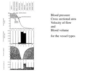
Vein structure and function • Veins :Thin, elastic muscle layer with semilunar valves that prevent the blood from flowing in the opposite direction. • Veins carry deoxygenated blood (with the exception of pulmonary veins and umbilical vein).
Artery structure and function • Arteries :Thick, elastic muscle layer that can handle high pressure of the blood flowing through the arteries. • Arteries carry oxygenated blood (with the exception of the pulmonary artery and umbilical artery).
Capillary structure and function • Capillaries: Very thin walls made up of endothelial cells, which allow substances to move through the wall with ease. • They are the site where oxygen and other nutrients in the blood are delivered to the tissues of the body
VIDEO!!!! • http://www.youtube.com/watch?v=SvAVu-7E2gA