Download
1 / 19
200 likes | 323 Vues
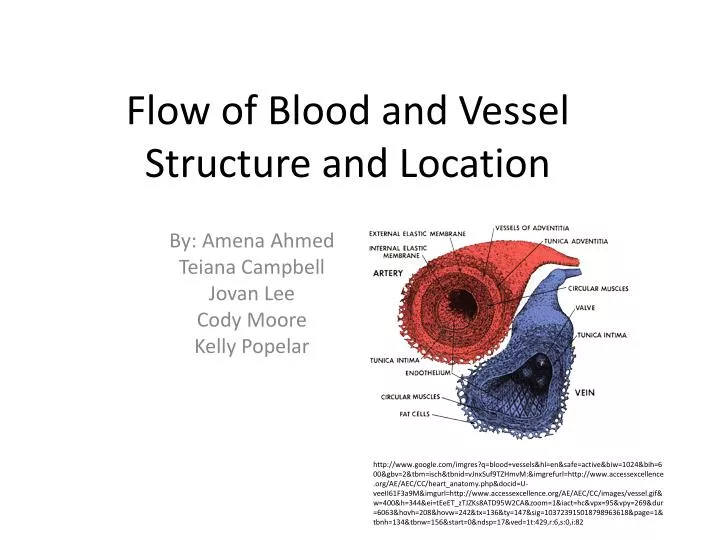
Flow of Blood and Vessel Structure and Location. By: Amena Ahmed Teiana Campbell Jovan Lee Cody Moore Kelly Popelar.

E N D
Flow of Blood and Vessel Structure and Location By: Amena Ahmed Teiana Campbell Jovan Lee Cody Moore Kelly Popelar http://www.google.com/imgres?q=blood+vessels&hl=en&safe=active&biw=1024&bih=600&gbv=2&tbm=isch&tbnid=vJnxSuf9TZHmvM:&imgrefurl=http://www.accessexcellence.org/AE/AEC/CC/heart_anatomy.php&docid=U-veeII61F3a9M&imgurl=http://www.accessexcellence.org/AE/AEC/CC/images/vessel.gif&w=400&h=344&ei=tEeET_zTJZKs8ATD95W2CA&zoom=1&iact=hc&vpx=95&vpy=269&dur=6063&hovh=208&hovw=242&tx=136&ty=147&sig=103723915018798963618&page=1&tbnh=134&tbnw=156&start=0&ndsp=17&ved=1t:429,r:6,s:0,i:82
Major Veins and Arteries that Lead to and from the Heart • Left Coronary Artery- supplies blood to the left ventricle, left atrium, and interventricular septum • Branches into the marginal and posterior interventricular • Right Coronary Artery- supplies blood to the right atrium and to portions of both ventricles • Branches into the circumflex and anterior interventricular • Small tributaries from these branches of the left and right coronary arteries form interconnections called anastomoses
Major Veins and Arteries Continued… • Great Cardiac and Middle Cardiac Veins carry blood away from the coronary capillaries and drain into the coronary sinus (a large, thin-walled vein in the posterior portion of the coronary sulcus)
Major Veins and Arteries Found on the Heart • Right Coronary Artery(Marginal branch and posterior interventricular branch) • Anterior Cardiac Veins • Small Cardiac Vein • Left Coronary Artery (Circumflex branch, anterior interventricular branch, and posterior left ventricular) • Great Cardiac Vein • Middle Cardiac Vein • Posterior Cardiac Vein
Major Veins and Arteries Found on the Heart http://www.google.com/imgres?q=veins+and+arteries+on+the+heart&hl=en&safe=active&gbv=2&biw=1024&bih=600&tbm=isch&tbnid=cAYwU2vQEnw4-M:&imgrefurl=http://www.centurahealthinfo.org/In-Depth%2520Reports/10/000003.htm&docid=tHqRXoVHASOgQM&imgurl=http://www.centurahealthinfo.org/graphics/images/en/1097.jpg&w=400&h=320&ei=e0uET7GnCYyk8gSgwey3CA&zoom=1&iact=hc&vpx=191&vpy=138&dur=2531&hovh=201&hovw=251&tx=114&ty=109&sig=103723915018798963618&page=1&tbnh=122&tbnw=153&start=0&ndsp=18&ved=1t:429,r:1,s:0,i:69
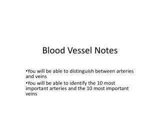
Vein Structure and Function • Veins carry blood to the heart • Inferior Vena Cava (Carries blood from body below the heart) and Superior Vena Cava (Carries blood from body above the heart) • Left and Right Pulmonary Veins- oxygen rich blood drains from the lungs and is returned to the left side of the heart through the 4 pulmonary veins (2 left, 2 right)
Vein Structure and Function Continued… • Right Pulmonary Vein – carry blood from the right lung into the left atrium through the left ventricle and out of the aorta • Left Pulmonary Vein – carry blood from the left lung into the left atrium through the left ventricle and out of the aorta
Vein Structure and Function http://www.google.com/imgres?q=vein+structure&hl=en&safe=active&gbv=2&biw=1024&bih=600&tbm=isch&tbnid=QoouXkC2Pv0TnM:&imgrefurl=http://legacy.owensboro.kctcs.edu/gcaplan/anat2/notes/APIINotes5%2520Circulatory%2520Anatomy.htm&docid=wC7NEnxxSCw4YM&imgurl=http://legacy.owensboro.kctcs.edu/gcaplan/anat2/notes/0672l.jpg&w=640&h=480&ei=4UuET-DpAYys8ASrnIC1CA&zoom=1&iact=hc&vpx=88&vpy=280&dur=3672&hovh=194&hovw=259&tx=200&ty=118&sig=103723915018798963618&page=1&tbnh=119&tbnw=158&start=0&ndsp=18&ved=1t:429,r:6,s:0,i:80
Capillary Function • Only blood vessels whose walls permit exchange between blood and the surrounding interstitial fluid • Capillaries do not function individually, they function in a network called a capillary bed • Diffuses water, small solutes, and lipid-soluble materials into the interstitial fluid • In some places such as the brain and kidneys, larger proteins are able to go through the capillaries
Capillary Function Continued • Blood goes to venules • Alternate routes for blood flow are formed by anastomosis (outlet) • Sometimes blood bypasses capillaries and goes through arteriovenous anastomosis, which is a arteriole connected to a venule • If one capillary is blocked, others continue to work and supply blood
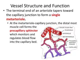
Capillary Structure • Capillary walls are extremely thin • Consists of a single layer of endothelial cells inside the membrane • Very small diameter, so blood flow is slow • Diameter is relatively close to that of a red blood cell • Entrance is guarded by a precapillary sphincter which is a band of smooth muscle • The smooth muscle contracts and narrows the diameter of the capillary opening, allowing the blood to enter into the capillary
Capillary Structure m/imgres?q=capillaries&um=1&hl=en&safe=active&sa=N&rls=com.microsoft:en-us:IE-SearchBox&biw=1024&bih=600&tbm=isch&tbnid=FiftVhhaE0bxbM:&imgrefurl=http://faculty.stcc.edu/AandP/AP/AP2pages/Units18to20/vessels/capillar.htm&docid=94xKhG3wubtoQM&imgurl=http://faculty.stcc.edu/AandP/AP/imagesAP2/bloodvessels/endothelia.jpg&w=366&h=376&ei=sUWET9nbC4WK8QTjy62gCA&zoom=1&iact=rc&dur=63&sig=109636002017002052041&page=1&tbnh=127&tbnw=124&start=0&ndsp=16&ved=1t:429,r:7,s:0,i:84&tx=86&ty=106
Artery Structure and Function • Carry blood away from the heart and toward a peripheral capillary • Within the pulmonary trunk the blood flows into the left and right pulmonary arteries • Arteries branch off repeatedly and gradually decrease in size until they become arterioles • Arterioles- the smallest vessels of the arterial system
Artery Structure and Function Continued… • Types of Arteries – Elastic, muscular, and arterioles • Elastic- large, extremely resilient vessels with diameters up to 2.5cm/1 in • Example: Pulmonary and aortic trunks • Thin walls, contain a tunica media dominated by elastic fibers rather than smooth muscle cells, able to absorb pressure shock
Artery Structure and Function Continued… • Muscular- AKA medium sized/distribution – distribute blood to peripheral organs • Example: Neck arteries • The tunica media contains more smooth muscle and fewer elastic fibers • Arterioles- smaller than muscular arteries • Has one of three layers of smooth muscle that enables muscular arteries and arterioles to change their diameter
Blood Flow Though the Body • Superior cava- gives blood to the head chest, upper extremities, Upper body • Inferior Cava- gives blood to the Lower Body • Pulmonary Circuit- a group of blood vessels that transports blood from exchange surfaces of the lungs • Systematic Circuit- Blood vessels that travel through the rest of the body • *the systematic veins must travel through the pulmonary system before restarting the cycle to collect oxygen and remove carbon dioxide
Blood Flow Through the Heart • Right Atrium- receives blood from the systematic surface • Right Ventricle- receives blood from the right atrium through openings called cusps • Tricuspid valve- Contains the cusps, pupillary muscles on the right ventricle that limit the movement of cusps and flow of blood • Pulmonary Semilunar Valve- after blood flow through the pulmonary trunk, this valve controls blood flow to the left and right pulmonary arteries • *oxygenated blood collects in the pulmonary veins then delivers it to the Left Atrium
Blood Flow Through the Heart Continued… • Bicuspid Valve- controls blood flow in this valve with two cusps, and tensing papillary muscles • Aorta- blood continues into the Aortic Semilunar Valve then the Aorta to start the systemic circuit
Blood Flow Through the Heart http://www.google.com/imgres?q=blood+flow+through+the+heart+diagram&hl=en&safe=active&gbv=2&biw=1024&bih=600&tbm=isch&tbnid=S2ElgCPYmnfWTM:&imgrefurl=http://en.wikibooks.org/wiki/Anatomy_and_Physiology_of_Animals/Cardiovascular_System/The_Heart&docid=0ko8BTbCW0WCyM&imgurl=http://upload.wikimedia.org/wikipedia/commons/e/e2/Section_through_heart_to_show_valves_and_blood_flow.jpg&w=602&h=454&ei=L0yET8bTGYuy8QS8g-m1CA&zoom=1&iact=hc&vpx=87&vpy=134&dur=2750&hovh=195&hovw=259&tx=133&ty=100&sig=103723915018798963618&page=1&tbnh=118&tbnw=156&start=0&ndsp=18&ved=1t:429,r:0,s:0,i:67