Download
1 / 25
250 likes | 1.23k Vues
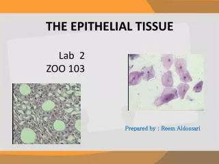
Introduction to Histology Tissues of the Body Epithelial Tissue and Glands. GEMP – 2006 Avinash Bharadwaj. Histology – What and Why. Histology – Study of Tissues (histos = web/tissue) By extension, microscopic structure of the body “Basic Histology” – study of (primary) tissues

E N D
Introduction to HistologyTissues of the BodyEpithelial Tissue and Glands GEMP – 2006 Avinash Bharadwaj
Histology – What and Why • Histology – Study of Tissues (histos = web/tissue) • By extension, microscopic structure of the body • “Basic Histology” – study of (primary) tissues • “Systemic Histology” – Organs and Systems • Also called microanatomy. • Histology • Basis of function • Stepping stone to cellular basis of disease – histopathology
A Different World…! • Microscopes – many varieties • Special preparation of material • Largely two-dimensional • Interpretation, analysis and application • Histological scale • Unit of measurement – Micron/micrometre (μ or μm) = 1/1000 mm or 10-6 m • Most material is in the form of sections (slices), usually 4 to 8 μ thick. • Thinner sections for electron microscopy
The Cell Details of cell biology and cell division are being covered elsewhere. If there are specific histology-related questions feel free to ask me! • The unit of life • Cell membrane • Cytoplasm • Organelles and their functions – emphasis on mitochondria, rough and smooth endoplasmic reticulum, Golgi complex, membrane-bound vesicles, lysosomes. • Nucleus • Nuclear membrane • Chromosomes – seen as such only during cell division • Chromatin • Euchromatin Lighter areas – active, uncoiled DNA • Heterochromatin Darker areas – compact material • A euchromatic nucleus is large, round and pale – active cell
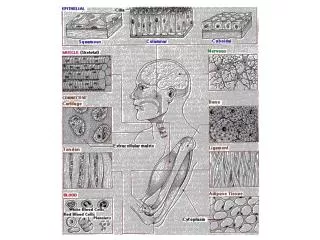
Primary (Basic) Tissues • Tissue – a group of cells serving a common function. • Immense variety of body structures all boil down to four principal tissue types • Epithelium, muscle, nervous tissue, connective tissue. (In histological technique the term tissue is used to mean any bit of body structure to be processed for microscopic examination).
Primary (Basic) Tissues • Epithelium • Covers body surfaces, lines organs and cavities. • Muscle • Sliding protein filaments – movement. (Sliding protein filaments also the basis of movement at cellular level. • Nervous tissue • Generation and transmission of electrical changes in cell membranes. • Supporting cells also included. • Connective tissue • Binding and packing material – mechanical strength, elasticity. • Other types of support – carrying nerves and blood vessels • Wide variety of functions.
Cavity (lumen) with food. • Epithelium for lining secretion and absorption. • Muscle for moving food. • Connective tissue to join. • Nerves (not seen). Integration of Tissues Small intestine as an example.
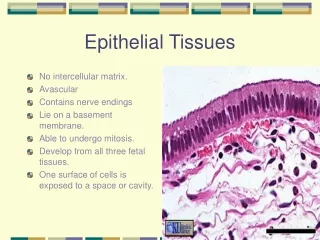
Epithelium • Covering – external surface • Epidermis of the skin • Cornea of the eye • Hair and nails are epidermal structures • Lining cavities • Internal organs – intestines, other tubular structures including heart and blood vessels. • “Body cavities” • Characteristic structural features • Compact sheets of cells – ‘tailored’ into a variety of shapes • Very little intercellular substance compactness • Cell junctions integrity of sheets, cell-to-cell communication and other functions. • Basement membranes • Avascularity … supporting tissue required.
Epithelium • Barrier between different environments. • Selective passage of substances across an epithelium – variety of mechanisms : diffusion, active transport, membrane-bound ‘vesicles’. • Body and the exterior. • Intestine – selective absorption of contents. • Secretion and absorption • Special functions • Protective – barrier, thick covering. • Sensory • Some functions demand a single layer of cells – ‘simple’ epithelium. • Friction/wear and tear : Multiple layers of cells – stratified epithelium.
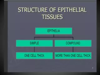
Epithelium – Classification • Simple epithelia named by shapes of cells. • Flat cells – squamous • Tall cells – columnar • Internediate – cuboidal • Stratified epithelia named by shapes of surface cells. • Stratified squamous • Stratified cuboidal • Stratified columnar • “Transitional” (surface cells can change their shapes). Stratified cuboidal and columnar epithelia : few examples. “Transitional” epithelium is found in the urinary system – it will be studied later!
Epithelia as sheets Simple squamous Simple cuboidal Simple columnar Epithelium as a tube Stratified squamous, keratinised Stratified squamous – nonkeratinised Nucleated, flat surface cells
Simple Squamous Epithelium • Single layer of flat cells facilitates transport across cells. • Blood vessels. Allows exchanges across capillary walls, also maintains a smooth surface in the heart and other vessels. The latter is important as blood tends to clot on rough surfaces. • Lungs – gas exchange across epithelium. Air-containing spaces and blood capillaries are both lined by this epithelium. The blood-air barrier is thus made of two epithelial layers and some very fine connective tissue between the two. • There are a few other sites where this epithelium is seen. • These will be discussed with the relevant organs/systems. • Also bear in mind that even for the sites mentioned above, the • role of the epithelium will be discussed further when these • systems are studied.
Secretory “vesicles” Absorption Microvilli (Brush Border) Golgi rER Simple Columnar Epithelium Cells of simple columnar epithelium show specialisations suited to function. Secretion Individual microvilli are too fine to be seen with the light microscope. With the LM, one sees only a pink band on the surface
Stages in the functional cycle of a mucus producing goblet cell. Simple Columnar Epithelium • Cilia • Hairlike, motile projections. • Each cilium anchored by a basal body. • For movement of fluid on the surface. • Fluid may be watery or viscous (mucus). • Cilia “beat” in one direction.
Stratified (Compound) Epithelia • Two or more (usually) layers (strata) • Basal layer : rests on the basement membrane. • Surface layer faces outward or faces the cavity. • Named by the appearance of the surface cells.
Stratified Squamous Epithelium In both these epithelia, the basement membrane is shown as a brown line. As the surface cells are ‘rubbed off’ or shed, the cell numbers are maintained by division in the basal layer. Note that on the left, the cells at the surface are nucleated – live. On the right, the surface layers are made of dead cells. The dead cells form the keratin layer – a complex protein-lipid layer which is relatively waterproof. A keratinised surface is a dry surface (except when there is excessive sweating!). Note the most superficial living cells – loaded with granules. These granular cells herald keratinisation. A non-keratinised surface is a ‘wet’ surface. Where does one expect to find these epithelia?
Other Stratified Epithelia • Stratified cuboidal and columnar • Will be mentioned later. • “Transitional” • The shapes of surface cells change with degree of stretching. Also, the cells have specialised surface features. • This is found in the urinary system and will be studied later.
Pseudostratified Columnar Epithelium. • Two or more cell types. • Small, undifferentiated cells. • Tall columnar differentiated cells. • All cells touch basement membrane. • False stratified appearance • Nuclei at different levels. • With ciliated cells • Nonciliated cells produce watery fluid. Uterine tube. • With ciliated cells and goblet cells. • Trachea and other respiratory passages.
Glands • Epithelial specialisation for secretion. • A patch of epithelial cellsOrA downgrowth that proliferates. • Microscopic, part of an organOrA large organ by itself. • In either case, elements of supporting connective tissue exist.
Ep Cavity 1 3 4 2 Gland – From an Epithelium A portion of an epithelium grows into the underlying supporting connective tissue. The downgrowth develops into a secretory portion and a duct. This is an exocrine gland. If the duct disappears, an extensive capillary network collects the secretions in an ‘endocrine’ Gland (4).
Types of Glands Glands can be classified in may overlapping ways. • Exocrine (with ducts) and endocrine (ductless). • Exocrine glands : • Simple (single duct) and compound (branched duct system). • Type of secretion – serous, mucous or mixed. • Mode of secretion – extent of cytoplasmic loss. Rather than making it a learning issue, it is fruitful to understand these terms as we come across them. Besides, there are glands that defy the concepts of classification! The following diagram serves to illustrate some of the types of glands. Some of the terms are purely descriptive!
A is a ‘unicellular’ gland (a goblet cell!). B and C may be ignored! – They represent glands with no specialised duct portion. D is a simple tubular gland. E is a simple, coiled tubular gland. In F and G, the duct is single, but the secretory portions are branched. H is an unusual gland of a type found in the eyelid. J and K are compound glands. Round secretory units are described as acini or alveoli. It is also possible to see ‘tubuloacinar’ secretory portions.
Cytoplasmic Loss • Merocrine (eccrine) gland • Little or no loss of cytoplasm. • Apocrine gland • Partial loss of (apical) cytoplasm. • Holocrine gland • Entire cell disintegrates to release secretion. These terms are best understood in the context of glands of the skin.
Serous and Mucous Glands • A glandular unit with serous secretions shows round nuclei, well-stained cytoplasm and a small lumen. Usually the basal portion of the cytoplasm is basophilic due to rER, the apical portion with secretory granules is acidophilic. • A mucous unit has cells loaded with lightly stained mucus in the cytoplasm. This pushes the nuclei towards the outer side.
Larger Glands • A larger gland is histologically a complex three-dimensional structure with secretory units and branches of ducts sectioned in various planes. These details will be studied with the digestive system where we come across a variety of glands. • It is, however, worth noting that such a proliferation of epithelial cells compresses the connective tissue. Such partitions of connective tissue separate parts of glands of varying sizes, called lobes and lobules. The connective tissue framework is called the stroma, the epithelial element is the parenchyma. These terms are met with in many solid structures. Last slide!