Download
1 / 65
720 likes | 982 Vues
NEUROANATOMY Lecture : 1 Introduction and Organization of the Nervous System, Anatomy of the Spinal Cord. Prepared and presented by: Dr. Iyad Mousa Hussein, MD, Ph.D in Neurology Head of Neurology Department Nasser Hospital. LECTURE OBJECTIVES:.

E N D
NEUROANATOMY Lecture : 1 Introduction and Organization of the Nervous System, Anatomy of the Spinal Cord Prepared and presented by: Dr. Iyad Mousa Hussein, MD, Ph.D in Neurology Head of Neurology Department Nasser Hospital
LECTURE OBJECTIVES: The Main Structures and Functions of the Nervous System. The Development of the Nervous System. Structures and Classification of Neurons. Types of Neuroglial Cells. Shape, Length, Diameter, Location, Beginning and Termination of the Spinal Cord. Functions and Meninges of the Spinal Cord. External Structure of the Spinal Cord. The Spinal Cord Segments. Relationship Between the Spinal Cord Segments and the Vertebra. The Internal Structure of the Spinal Cord. Blood Supply and Venous Drainage of the Spinal Cord.
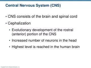
Introduction of the Nervous System Nervous system (NS) is the system of communication, the aim of communication is to keep body homeostasis, NS does its function through: 1. Detection of the changes. 2. Evaluation of the information. 3. Responding properly. 4. Coordination voluntary and involuntary behaviors. 5. Higher order cognition.
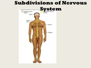
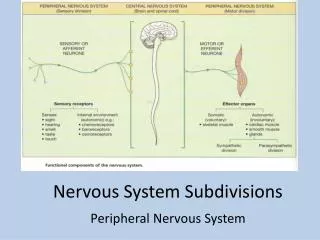
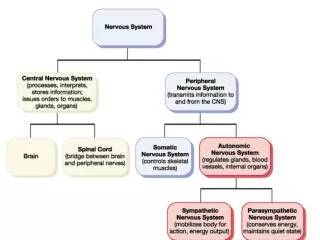
Classification of the Nervous System • A. According to the location: • 1. Central nervous system (CNS): • a. Brain (intracranial part). • b. Spinal cord (extracranial part). • 2. Peripheral nervous system (PNS): • a. Cranial nerves. • b. Spinal nerves. • B. According to the function: • 1. Somatic (voluntary or craniospinal) NS: • a. Motor NS. • b. Sensory NS • 2. Autonomic NS: • a. Sympathetic NS. • b. Parasympathetic NS. • The somatic nervous system supplies the skeletal muscles, while the autonomic nervous system supplies the smooth and cardiac muscles.
The Development of the Nervous System The Entoderm: gives rise to the gastrointestinal tract, the lung, and the liver. The Mesoderm: gives rise to the muscles, connective tissue, and the vascular system. The Ectoderm: gives rise to the nervous system. During the third week of development, the ectoderm → Neural plate → Neural groove → Neural tube.
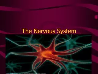
The Neurobiology of the Neurons Neuron:is the name give to the nerve cell and all its processes. Each neuron consist of: 1. Cell body. 2. Axon. 3. Dendrites.
Bipolar Neuron Unipolar Neuron Multipolar Neuron
Classification of Neurons According to the Function 1. Motor neuron. 2. Sensory neuron. Structure of the Nerve Cell Body 1. Nucleus. 2. Cytoplasm: it is formed of the following structures: a. Nissl substance. b. The Golgi complex. c. Mitochondria. d. Microfilaments. e. Microtubules. f. Lysosomes. g. Centrioles. h. Lipofuscin, Melanin, Lipid, and glycogen. 3. Plasma membrane.
Types of neuroglial cells The neurons of the CNS are supported by several varieties of nonexcitable cells, which together are called neuroglia. Types of neuroglial cells: 1. Astrocytes. 2. Oligodendrocytes. 3. Microglial cells. 4. Ependymal cells.
The Spinal Cord The spinal cord (SC) is an extracranial part of the central nervous system enclosed inside the vertebral column.
The Spinal Cord Shape:compressed cylindrical column. Number of neurons in human SC:about 13,500,000 neurons. Length of human SC: about 45 cm (male); 43 cm (female). Weight of human SC: about 35 g. Diameter: about 1 cm. Location:It is located in the spinal (vertebral) canal.
Beginning of the Spinal Cord The spinal cord is continuation of the medulla oblongata which begin from the foramen magnum a downward. • Termination of the Spinal Cord • At the third month of intrauterine life: • the SC fills the whole spinal canal. • 2. At birth: it ends at the level of third lumbar vertebra. • 3. In the adult: it ends at the level of lower border of the first lumbar vertebra.
Functions of the Spinal Cord 1. Conduction:from and to the brain: a. Ascending tracts:conduct sensory impulses from SC to the brain. b. Descending tracts:conduct motor impulses from brain to the SC. 2. Reflexes center:for all spinal reflexes.
Meninges of the Spinal Cord • The spinalcord is covered by 3 membranes (meninges), • from inside to outside they are: • 1. Pia mater:it is closely adhesent to the spinal cord, ends at the lower border of the first lumbar vertebra. • 2. Arachnoid Mater: it lines the inner surface of the dura mater, ends at the level of the S2 vertebra. • 3. Dura mater:ends at the level of the S2 vertebra.
Epidural Space: it is located between the two layers of the dura mater. Subdural Space: it is located between the dura mater and arachnoid mater. Subarachnoid Space: it is located between the arachnoid and pia mater.
Anatomy of the Spine Length of human vertebral column:about 70 cm. Vertebrae: 7 Cervical 12 Thoracic 5 Lumbar 5 Sacral 3 Coccyx
Structure of the Vertebra 1. Vertebral body. 2. Lamina. 3. Two transverse processes. 4. Spinous process. 5. Superior and inferior articular processes 6. Spinal canal.
The Spinal Cord Segments There are 31 segments: 8 – Cervical segments. 12 – Thoracic (dorsal). 5 – Lumbar segments. 5 – Sacral segments. 1 – Coccygeal segments.
External Structure of the Spinal Cord The external surface of the spinal cord shows the following: 1. Anterior median sulcus (fissure). 2. Posterior median sulcus. 3. Right & left posterolateral (intermediate) sulci: at the line of attachment of the posterior (sensory) roots of the SC. 4. Right and left anterolateral sulci: at the line of attachment of the anterior (motor) roots of the SC.
Enlargements of the Spinal Cord 1. Cervical enlargement:It is the C5, 6, 7, 8 and D1, 2 segments of the SC, corresponding to the region from which the brachial plexus arises → gives spinal nerves that supply the upper limbs. 2. Lumbar enlargement:It is the L1, 2, 3, 4, 5 and S1, 2 segments of the SC, corresponding to the region from which the lumbar and sacral plexuses arises → gives spinal nerves that supply the lower limbs.
Epiconus: It is the L4, 5 and S1, 2 segments of the SC. Conus medularis: It is the S3, 4, 5 segments of the SC. The Cauda Equina: It is located below the lower border of the first lumbar vertebra, the spinal canal is filled by the collection of lumbo-sacral roots which descends in this space to their corresponding intervertebral canal.
The Relationship Between the Spinal Cord Segments and Vertebra
The Roots of the Spinal Cord • Two posterior (dorsal) roots: there are sensory fibers (afferent fibers) → 31 pairs of posterior roots (right and left). • 2. Two anterior roots: there are motor and autonomic fibers (efferent fibers) → 31 pairs of anterior roots (right and left). • The anterior and posterior roots will join to form a single mixed nerve called the spinal nerve→ 31 pairs of spinal nerves (right and left). • Each spinal nerve emerges from the intervertebral foramen.
The Spinal Nerves • There are 31 pairs of spinal nerves are • attached to the SC: • 8 – Cervical nerves. • 12 –Thoracic (Dorsal) nerves. • 5 – Lumbar nerves. • 5 – Sacral nerves. • 1– Coccygeal nerve.
Internal Structure of the Spinal Cord A transverse section in the SC is formed of: 1. Outer white matter. 2. Inner grey matter. 3. Central canal (1 mm in diameter): is present in gray matter.