Download
1 / 5
50 likes | 112 Vues
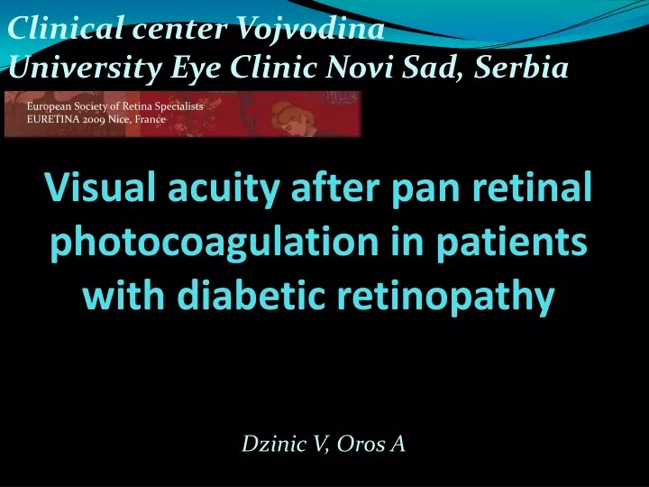
Visual acuity after pan retinal photocoagulation in patients with diabetic retinopathy. Dzinic V, Oros A. Clinical center Vojvodina University Eye Clinic Novi Sad, Serbia. European Society of Retina Specialists EURETINA 2009 Nice, France. Purpose:. Material and methods:.

E N D
Visual acuity after pan retinal photocoagulation in patientswith diabetic retinopathy Dzinic V, Oros A Clinical center Vojvodina University Eye Clinic Novi Sad, Serbia European Society of Retina Specialists EURETINA 2009 Nice, France
Purpose: Material and methods: • 35 patients (69 eyes), were treated by pan retinal laser photocoagulation (PRP) • pause between sessions was one week. • all patients used local therapy of NSAID during treatment, and one week after last session. • VA was measured by Snellen chart, before treatment, during second and fourth week and three months after treatment to examine effect of pan retinal laser photocoagulation (PRP) on visual acuity (VA) in patients with severe non-proliferative diabetic retinopathy, without clinically significant macular edema. (CSME)
Results: Average VA before LFK was 0.568±0.34 During follow up period, in the first month, VA was decreased gradually
Results: Average VA after three months of treatment was 0.6±0.33 (p>0.05).
Conclusion: diabetic retinopathy impairs macular function, despite absence of CSME by destroying peripheral retina PRP has beneficial effects on improving metabolic status of macula better perfusion leads to improvement in retinal function and respectively visual acuity