Download
1 / 9
100 likes | 215 Vues
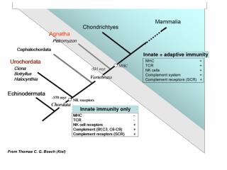
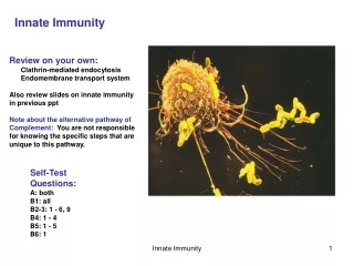
Innate immunity mediated by APOBEC3G.

E N D
Innate immunity mediated by APOBEC3G Expression of APOBEC3G in the virus-producing cell (left) can lead to abortive retroviral infection of the target cell (right). APOBEC3G is incorporated into progeny virions as they assemble and bud. After entry of the viral core into the target cell, the viral RNA genome (red) is reverse transcribed into minus-strand cDNA (light blue). APOBEC3G triggers dC to dU deamination of the minus-strand cDNA (probably some time after degradation of the plus strand RNA by RNase H; not shown). The dU residues in the first-strand cDNA may then lead to excessive hypermutation capable of inactivating viral functions (left); degradation mediated by base-excision-repair enzymes (middle); or misreplication of the second cDNA strand. HIV Vif can counteract APOBEC3G-mediated immunity by binding to APOBEC3G and inducing its ubiquitination anddegradation. Nature Immunology 4, 641-643 (2003)
Figure 1. DNA Deamination by CEM15/APOBEC3G. (A) Deamination of dC within a single-stranded oligonucleotide by purified His-tagged CEM15/APOBEC3G (as well as by recombinant APOBEC1 control) was monitored by subsequent treatment of the oligonucleotide with uracil-DNA glycosylase (UDG)/NaOH which breaks the oligonucleotide at the site of dC to dU deamination (illustrated to the left of the gel image). (B) Western blot of E.coli extracts expressing recombinant CEM15/APOBEC3G (upper band, lane 1) or a control plasmid (lane 2).
Figure 2. CEM15/APOBEC3G Diminishes MLV Infectivity. 293T-derived stocks of YFP-encoding MLV produced by cotransfection with a control vector or with 1 mg or 3 mg of pCEM15:HA were used in challenges of Mus dunni fibroblasts across a range of input inocula (normalized units of RT). Similar data were obtained in parallel challenges of N-3T3 cells (data not shown).
Figure 3. Mutation of Retroviral First-Strand cDNA by CEM15/APOBEC3G. (A) GFP fluorescence of target 293T cells 48h after challenge with equivalent levels of MLV-GFP virions derived from 293T cells stably expressing CEM15/APOBEC3G or a control plasmid. The percentages of cells within the GFP-positive window are indicated. (B) Profiles of the relative positions of the G to A transition mutations apparent in 12 representative 730 bp GFP sequences derived from GFP-lo and GFP-hi populations of cells challenged with MLV-GFP grown on CEM15/APOBEC3G-expressing cells or nonexpressing controls. Two independently derived CEM15/APOBEC3G+ clones (and two independent controls) were analyzed for each experiment (separated by a space). The G to A transitions are depicted by vertical lines whereas other single nucleotide substitutions (20 in total) are indicated by lollipops and thick, horizontal black bars represent the two deletions detected. Note that each pair of panels (lo and hi) represents sequences recovered from 293T target cells, but the viral stocks used in each experiment (each panel pair) were derived from independent clones either expressing or not expressing CEM15/APOBEC3G. (C) A comparison of the extent of GFP mutation in the different samples. The pie charts depict the proportion of MLV sequences carrying the indicated number of mutations within the 730 bp interval sequenced. The total number of sequences determined in each data set is indicated in the center.
Figure 4. Retroviral cDNA Mutation Spectra. (A) Mutations detected amongst GFP sequences amplified from GFP-lo enriched target cell populations. Mutations derived from MLV-GFP challenged target cells in which the viral stocks used were grown in CEM15/APOBEC3G-expressing cells are depicted above the 730 bp consensus (the viral plus or coding strand is shown) and those derived from vector-expressing cells are shown below. Deletions are boxed. (B) GFP mutations from GFP-hi enriched cell populations.
Figure 5. Nature and Local Preferences of CEM15/APOBEC3G Mutation. Nucleotide substitution preferences for the entire set of GFP mutations detected in target cells infected with MLV-GFP that had been grown on a CEM15/APOBEC3G-expressing producer cells in comparison to those detected in target cells infected with MLV-GFP grown on a vector-expressing producer cells. (B) Delineation of the preferred local sequence context for CEM15/APOBEC3G-mediated dC deamination in MLV-GFP. All 734 mutated positions were aligned with respect to the dC residue targeted for deamination on the minus (first)-strand cDNA and the frequency (as a percentage) with which each of the four bases is found at adjacent positions was calculated.
Figure 6. CEM15/APOBEC3G Is Packaged into MLV Particles and HIV Vif Is Able to Inhibit Its Function. YFP-encoding MLV virions were purified from cells (Figure 2) that did not (left lane) or did express CEM15/APOBEC3G (right lane) and analyzed by immunoblotting using antibodies specific for MLV Gag or CEM15/APOBEC3G (HA epitope). (B) HIV Vif diminishes CEM15/APOBEC3G-mediated immunity to YFP-encoding MLV as monitored using the transient expression system (as in Figure 2 ) in the presence of pCEM15:HA (3 mg) with or without pcVIF (1 mg). (C) Restoration of infectivity of CEM15/APOBEC3G-exposed MLV-GFP by expression of HIV Vif during viral stock production from 293T cells stably expressing CEM15/APOBEC3G (as in Figure 3A). The marginal restoration of infectivity in experiment 2 correlates with the approximately 3-fold higher expression of CEM15/APOBEC3G in this cell line over that of the cells used in experiment 1.
Figure 7. Mechanism of CEM15/APOBEC3G Triggered Innate Immunity. Two possible models are contrasted, one illustrating viral inactivation by excessive mutation (left; dU is recognized as dT by DNA polymerases) and the other envisioning viral destruction being furthered by recognition of uracil in DNA (right; a development of a previous speculation; Harris et al., 2003). Deoxyuridine in the first-strand cDNA would be a target for excision by uracil-DNA glycosylase, generating an abasic site (asterisked), and therefore a probable target for endonucleolytic cleavage. However, the identity of this endonuclease is uncertain but is presumed to be analogous to the apurinic/apyrimidinic endonucleases that act on abasic sites in dsDNA in the base excision repair pathway (Lindahl and Wood, 1999). A third possibility suggested by the results of Klarmann et al. (2003) is that the presence of deoxyuridine in minus (first)-strand cDNA may alter the specificity of initiation of plus (second)-strand cDNA synthesis. RTase is reverse transcriptase.