Download
1 / 32
320 likes | 429 Vues
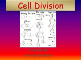
Cell Division. “ Omnis cellula e cellula ”. The Key Roles of Cell Division. Essential for perpetuation of life: Reproduction of unicellular forms Development, Growth, & Repair of multicellular forms. DNA – The Blue Prints for Life. Genome – cell’s endowment of DNA

E N D
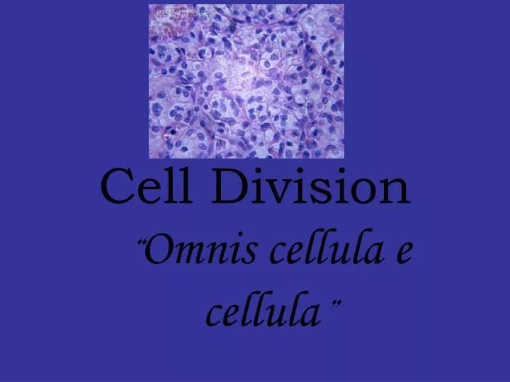
Cell Division “Omnis cellula e cellula”
The Key Roles of Cell Division • Essential for perpetuation of life: Reproduction of unicellular forms • Development, Growth, & Repair of multicellular forms
DNA – The Blue Prints for Life • Genome – cell’s endowment of DNA • Chromatin DNA-protein complex • Chromosomes “colored bodies” make it possible for 3 meters of DNA to fit in one eukaryotic cell • Chromosome # is species specific • Somatic cells vs gametes • Sister chromatids duplicated chromosomes • Centromere • Telomere
The Mitotic Cycle • Interphase 90% • G1 = Grow, normal fx. • S = Synthesis of DNA • G2 = Prepares for cell division • Mitosis • PMAT • Cytokinesis –cell pinches in 2 • Animals: Cytoplasm side consists of a ring of actin-myosin filaments that move past each other causing the ring to contract (drawstrings) • Plants: Golgi Vesicles move to middle of cell coalesce cell plate
Interphase • Nucleus is well defined in a nuclear envelope • DNA is in the form of loosely packed chromatin fibers • Accounts for 90% of cell cycle • Cell grows and copies DNA in preparation for mitosis
Prophase • Chromatin fibers become tightly coiled into discrete chromosomes • The nucleoli and nuclear envelope begin to disappear • Each duplicated chromosome appears as two identical sister chromatids joined together • Mitotic spindles begin to form from microtubules extending from centrosomes • Kinetochore forms at centromere region
Metaphase • Centrosomes are at opposite poles of the cell • Chromosomes convene at metaphase plate • Kinetochores of the sister chromatids are attached to microtubules coming from opposite poles of the cell.
Anaphase • Paired centromeres of each chromosome separate, liberating the sister chromatids from each other • Kinetochore microtubules shorten – moving the daughter chromosomes to opposite poles of the cell.
Telophase • Daughter nuclei form at the two poles of the cell • Nuclear envelopes arise from the fragments of the parent cell’s nuclear envelope and other portions of the endomembrane system • Chromatin fibers of each chromosome become less coiled
http://www.cellsalive.com/cell_cycle.htm • http://www.cellsalive.com/mitosis.htm
Mitotic Spindle • Chromosome movement is controlled by mitotic spindle • Spindle arises from centrosomes • Kinetochore microtubules depolymerize, moving chromosomes throughout mitosis • Nonkinetochore microtubules elongate cell
Evolution of Mitosis(page 225) • Prokaryotes – daughter chromosomes move apart ???? • Dinoflagellates – nuclear envelope stays intact for chromosomes to attach; microtubules pass through n.e. reinforcing spatial orientation of nucleus fission • Diatoms – n.e. remains intact; microtubules from a spindle w/i nucleus separating the chromosomes nucleus splits • Eukaryotes – n.e. breaks down spindle fibers form outside of nucleus attach to kinetochore for separation of sister chromatids
Regulation of the Cell Cycle • Cell Cycle Check points: G1, G2, & M • G1 checkpoint Most important; if cell does not receive signal to go ahead to S then cell will go into G0 (nondividing state: liver, neuron) • Cell cycle is driven by specific chemical signals present in cytoplasm (not dominoes) • Cyclin = protein that has fluctuating levels during cell cycle; synthesized during interphase • Cdk’s- protein kinases that must attach to cyclin to be activated • MPF “maturation promoting factor”: composed of Cdk & cyclin complex; promotes mitosis by phosphorylating various proteins • One indirect effect is the breakdown of its own cyclin (off switch)
Internal Signals: • M phase checkpoint Messages from kinetochores ensures that chromosomes are properly attatched to the spindle at metaphase. Why? • Protects against missing chromosomes • External Signals: Growth factors must be present to stimulate the growth of cells (specific) • Density dependent inhibition – growth factors and nutrients are insufficient to stimulate growth • Anchorage dependence – most animal cells must be anchored to a substratum to divide • CANCER CELLS EXHIBIT NEITHER OF THESE!!!!!
Cell Cycle Controland Mutation Controls in the Cell Cycle • Checkpoints exist in the cell cycle • Cell determines if cell is ready to enter next part of cell cycle http://highered.mcgraw-hill.com/olc/dl/120082/bio34a.swf
What Is Cancer? • Cancer begins when the proteins that regulate the cell cycle don’t work, the cell divides uncontrollably • Mutations can be inherited or induced by exposure to U.V. radiation or carcinogens that damage DNA and chromosomes
Cancer: Uncontrolled cell growth • Tumor • Malignant vs benign • Metastasis • Types of cancer • Carcinoma (epithelials) • Melanoma (melanocytes) • Sarcoma (muscle/connective) • Osteogenic (bone) • Leukemia (blood forming organs) ↑ WBC’s • Lymphoma (lymphatic) • Malignant cells trigger angiogenesis
Mutations to Cell-CycleControl Genes • Proto-oncogenes: Normal genes on many different chromosomes regulate cell division • When mutated, they become oncogenes • Many organisms have proto-oncogenes, so many organisms can develop cancer
Errors that cause cancer • p53 is a protein that functions to block the cell cycle if the DNA is damaged. If the damage is severe this protein can cause apoptosis (cell death). • p53 levels are increased in damaged cells. This allows time to repair DNA by blocking the cell cycle. • A p53 mutation is the most frequent mutation leading to cancer. • p27 is a protein that binds to cyclin and CdK blocking entry into S phase. Recent research (Nat. Med.3, 152 (97)) suggests that breast cancer prognosis is determined by p27 levels. Reduced levels of p27 predict a poor outcome for breast cancer patients.
From Benign to Malignant • Angiogenesis – growth of blood cells caused by secretions from cancer cells • Increases the blood supply to cancer cells: more oxygen and nutrients • Cancer cells can divide more • Tumors develop, sometimes filling entire organs
From Benign to Malignant • Contact inhibition in normal cells prevents them from dividing all the time, which would force the new cells to pile up on each other • Anchorage dependence in normal cells keeps the cells in place
Multiple Hit Model • Many changes, or hits, to the cancer cell are required for malignancy • Mutations can be inherited and/or can stem from environmental exposures • Knowledge of cancer risk factors is important • Earlier detection and treatment of cancer greatly increase the odds of survival
Detection Methods: Biopsy • Different cancers are detected by different methods, including high protein production possibly indicating a tumor • Biopsy, the surgical removal of cells, tissue, or fluid for analysis is performed • Under a microscope, benign tumors appear orderly and resemble other cells in the same tissue • Malignant tumors do not resemble normal tissue
5.6 Meiosis • Occurs within gonads (testes:ovaries) • Meiosis produces sex cells – gametes (sperm:egg) • Gametes have half the chromosomes (23) that somatic cells do (46) • Meiosis reduces the number of chromosomes by one-half
Meiosis contributes to Genetic Variation • There are millions of possible combinations of genes that each parent can produce because of: • Random alignment of homologous pairs • Crossing over • Random Feritlization (70 trillion)
Birth = paused at prophase I Puberty = finishes meiosis I Fertilization = finishes meiosis
*somatic cells *divide once diploid *forms identical cells *gametes *divide twicehaploid *forms different cells (crossing over) http://highered.mcgraw-hill.com/sites/0072437316/student_view0/chapter12/animations.html#
Conjoined Twins http://www.youtube.com/watch?v=ZzZYKggrB34&feature=fvsr http://www.youtube.com/watch?v=XM82Hs0LEpc