Download

1 / 79
990 likes | 2.46k Vues
JONCTIONS, ADHÉSION, MATRICE EXTRA CELLULAIRE. Plan. I – Jonctions cellulaires II – Adhésion cellulaire III – Matrice extra-cellulaire IV – Intégrines. III - LA MATRICE EXTRA CELLULAIRE. Définition. Tissu = cellules + espace extra cellulaire

E N D
Plan I – Jonctions cellulaires II – Adhésion cellulaire III – Matrice extra-cellulaire IV – Intégrines
Définition • Tissu = cellules + espace extra cellulaire • Rempli de macromolécules = matrice extra cellulaire • Protéines • Polysaccharides réseau en contact intime avec la surface des cellules
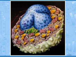
Fig 19-33 Cellules entourées de matrice extra cellulaire (bourgeon de membre)
Généralités • Jonctions tissus épithéliaux • Matrice extra cellulaire tissus conjonctifs • MEC > cellules • propriétés physiques des tissus • Quantités très variables • Cartilages, os +++ • Cerveau
Fig 19-34 Tissu conjonctif sous-jacent à un épithélium
Les différents types de tissus conjonctifs • Calcifiés : os, dents … • Transparents : cornée • Câble : tendon • Lame basale • ...
Rôles • Pendant longtemps : charpente inerte • Actuellement : actif et complexe régulation du comportement de la cellule en contact • Survie • Développement • Migration • Prolifération • Forme • Fonction
Propriétés • Composition moléculaire complexe • Incomplètement connue • Origine très ancienne • Présent dans tous les êtres pluricellulaires • Cuticule des vers et des insectes • Coquilles des mollusques • Parois des cellules végétales
Plan • Constituants du tissu conjonctif • Substance fondamentale • Glycosaminoglycannes • Protéoglycannes • Collagènes • Élastine • Fibronectine • Matrice extra cellulaire cytosquelette • Action de la cellule sur la matrice extra cellulaire : régulation de l’assemblage des fibrilles de fibronectine par les filaments d’actine intracellulaires • Action de la matrice extra cellulaire sur la cellule : guidage de la migration cellulaire par les glycoprotéines de la matrice • Lame basale • Matrice extra cellulaire comportement de la cellule • Dégradation de la matrice extra cellulaire et migration
3 - Collagènes • Les collagènes • La synthèse du collagène • Les maladies du collagène • Organisation des fibrilles de collagène
3 - Collagènes a) Les collagènes b) la synthèse du collagène c) Les maladies du collagène d) Organisation des fibrilles de collagène
a) - Les collagènes • Constituent une famille de protéines fibreuses • Rencontrée dans toutes les espèces animales • Protéine fibreuse • Sécrétée par les cellules du tissu conjonctif (et d’autres cellules) • Protéine la plus abondante dans la peau et l’os • Protéine la plus abondante des mammifères (25 % de la masse totale des protéines)
Jyrki HeinoThe collagen family members as cell adhesion proteinsBioEssays 29:1001-1010, 2007. • Review Article • Abstract • The collagen family of extracellular matrix proteins has played a fundamental role in the evolution of multicellular animals. • At the present, 28 triple helical proteins have been named as collagens and they can be divided into several subgroups based on their structural and functional properties. • In tissues, the cells are anchored to collagenous structures. Often the interaction is indirect and mediated by matrix glycoproteins, but cells also express receptors, which have the ability to directly bind to the triple helical domains in collagens. Some receptors bind to sites that are abundant in all collagens. However, increasing evidence indicates that the co-evolution of collagens and cell adhesion mechanisms has given rise to receptors that bind to specific motifs in collagens. These receptors may also recognize the different members of the large collagen family in a selective manner. • This review summarizes the present knowledge about the properties of collagen subtypes as cell adhesion proteins.
Caractéristiques de la molécule de collagène • Triple hélice : 3 chaînes enroulées • Très riche en proline et glycine • proline : structure en anneau stabilisation de l'hélice • glycine : • tous les 3 acides aminés • au centre de la chaîne • 3 acides aminés par tour
Chaîne alpha de la molécule de collagène • Une chaîne 1000 acides aminés • Hélice gauche • 3 acides aminés par tour • Un glycine tous les 3 acides aminés • -(Gly-X-Y)- • X souvent un proline • Y souvent un hydroxyproline
Fig 19-43 Structure d'une molécule de collagène (A) Chaîne (une sphère = un acide aminé) (B) 3 chaînes 1 chaîne 3 chaînes
Collagène • 25 chaînes différentes et 25 gènes • 50 exons par chaîne • 1 exon = 54 ou n X 54 nucléotides • 253 = 15 625 types de molécules ! • En fait qu ’une 20aine (28) • Les principaux :I, II, III, IV, XI … • Collagènes • fibrillaire (I, II, III, V, XI) (10-300 nm de diamètre) • associé aux fibrilles (IX, XII) • en réseau (IV, VII) • Transmembranaires
Collagènes • Fibrillaire : fibrilles fibres • Associé aux fibrilles • association des fibrilles les unes aux autres • et à d’autres éléments de la MEC • En réseau • IV : lame basale • VII : fibrilles d’ancrage de la lame basale au tissu conjonctif
Protéines collagène – like • XVII • possède un domaine transmembranaire • Composant des hémidesmosomes • XVIII • Lame basale des vaisseaux • Le clivage du domaine – C terminal endostatine
Endostatine • Résulte du clivage de l’extrémité – C terminale du collagène XVIII • Inhibe la formation de nouveaux vaisseaux • Étudiée comme drogue anti cancéreuse
Endostatins • Angiostatic proteins that are formed from proteolytic cleavage of COLLAGEN TYPE XVIII.
Physiological role of collagen XVIII and endostatin • Collagen XVIII/endostatin is a recently identified component of almost all epithelial and endothelial BMs. • This collagen is a heparan sulfate proteoglycan and contains • 10 collagenous (COL) domains • that are interrupted and flanked by noncollagenous domains (NC) • A proteolytic fragment of the C-terminal noncollagenous domain (NC1), termed endostatin, has been shown to have anti-angiogenic activity in vitro and in vivo.
Schematic representation of the two promoters and the splicing events giving rise to 3 different isoforms of COL18A1 transcripts. Alexander G. Marneros and Bjorn R. OlsenPhysiological role of collagen XVIII and endostatinThe FASEB Journal. 2005;19:716-728 • Exons 1-5 and the 3' exon 43 are shown. • Transcription from the upstream promoter and splicing of exons 1, 2, and 4 to exons 5-43 gives rise to the short isoform (NC11-303) • Transcription from the downstream promoter and splicing of exon 3 to exons 4-43 gives rise to the long isoform (NC11-728) • Transcription from the downstream promoter and splicing of the 5' portion of exon 3 to exons 4-43 gives rise to the intermediate isoform (NC11-493).
Alexander G. Marneros and Bjorn R. OlsenPhysiological role of collagen XVIII and endostatinThe FASEB Journal. 2005;19:716-728 • Schematic drawing of the interaction of collagen XVIII with other components of basement membranes (BMs) under endothelial or epithelial cells.
Abnormalities in the retinal pigment epithelium (RPE) and the retina in mice lacking collagen XVIII/endostatin in comparison to wild-type tissues. Alexander G. Marneros and Bjorn R. OlsenPhysiological role of collagen XVIII and endostatinThe FASEB Journal. 2005;19:716-728 • Basal laminar-like deposits in mutant mice are associated with • reduced content of RPE65 protein and retinyl esters in the retinal pigment epithelium (RPE), • reduced retinal rhodopsin content, • photoreceptor abnormalities, and increased expression levels of retinal GFAP.
Alexander G. Marneros and Bjorn R. OlsenPhysiological role of collagen XVIII and endostatinThe FASEB Journal. 2005;19:716-728 • In aged Col18a1–/– mice, pigmented macrophage-like "clump" cells migrate out of the iris toward the retina, where they may penetrate the inner limiting membrane (ILM). These cells accumulate in areas of increased retinal GFAP expression and photoreceptor disorganization.
Knobloch syndrome • The first indication that collagen XVIII/endostatin may be critical for the maintenance of ocular structures came from a linkage analysis of a consanguineous Brazilian family with Knobloch syndrome [MIM 267750]. • In this study the disease locus was mapped to the gene for collagen XVIII on chromosome 21q22.3 and a mutation within COL18A1 was identified. • Knobloch syndrome is • an autosomal recessive disorder characterized by the occurrence of • vitreoretinal degeneration with retinal detachment, • high myopia, • macular degeneration, • and occipital encephalocele. • Ocular abnormalities display • clinical variability • and may include congenital cataracts, iris abnormalities, or lens subluxation in some patients. • Besides the characteristic occipital encephalocele, further extraocular findings in Knobloch syndrome patients are rare and not typical of this syndrome. • However, the eye findings are severe and regularly lead to blindness at young age. • Family members of the consanguineous Brazilian family with Knobloch syndrome have a homozygous mutation at the AG consensus sequence at the 3' end of intron 1 in COL18A1, whereas obligate carriers of the disease allele are heterozygous for this mutation. • The mutation leads to skipping of exon 2 and the creation of a premature termination codon within exon 4 of the COL18A1 transcript.
Quelques types de collagène • Table 19-5
Génétique des collagènes • 25 chaînes différentes et 25 gènes • 50 exons par chaîne • 1 exon = 54 ou n X 54 nucléotides • • Naissance de ces collagènes par duplications multiples d’un gène primordial contenant 54 nucléotides et codant pour exactement 6 répétitions Gly – X – Y • (3-3-3) X 6 = 54 • [Gly-X-Y] [3 nucléotides - 3 nucléotides - 3 nucléotides]
Formules des collagènes • type I : [1(I)]2 2(I) • type II : [1(II)]3 • type III : [1(III)]3 • type IV : [1(IV)]2 2(IV) • type V : [1(V)]2 2(V) • ...
3 - Collagènes • a) les collagènes • b) la synthèse du collagène • c) Les maladies du collagène • d) organisation des fibrilles de collagène
(i) - Synthèse du collagène par les ribosomes • Ribosomes sur la membrane du réticulum endoplasmique • Synthèse de chaînes pro dans la lumière du réticulum endoplasmique • La chaîne pro possède • Le signal peptide à l’extrémité –N • Des acides aminés appelés propeptides aux deux extrémités de la chaîne
(ii) - Synthèse du collagène dans le réticulum endoplasmique • Certaines prolines et lysines hydroxyprolines et hydroxylysines • Certaines hydroxylysines sont glycosylées • Une chaîne se combine avec deux autres procollagène • Procollagène = triple hélice à liaisons hydrogène
Fig 19-45 Hydroxylysines et hydroxyprolines Rares dans les autres protéines animales Ces -OH liaisons hydrogène interchaînes stabilisation de la triple hélice
(iii) - Sécrétion du procollagène fibrillaire • Fusion des vésicules sécrétoires avec la membrane plasmique
(iv) – Transformation du procollagène fibrillaire en collagène • Excision des propeptides du procollagène fibrillaire par des enzymes protéolytiques en dehors de la cellule • Le procollagène devient collagène
(v) – Formation des fibrilles de collagène • Se fait donc dans l’espace extra cellulaire • Autoassemblage des molécules de collagène en fibrille : le collagène est 1 000 fois moins soluble que le procollagène • Les fibrilles se forment au contact de la membrane plasmique dans des invaginations profondes
Les pro-peptides • Guident la formation des molécules à 3 brins • Empêchent la formation de fibrilles dans la cellule (puisqu’ils sont retirés en dehors de la cellule)
Fig 19-44 Fibroblaste entouré de fibrilles de collagène dans le tissu conjonctif de la peau d'embryon de poulet Collagène fibrillaire
Collagène fibrillaire • Striation transversale de 67 nm en microscopie électronique
Explication moléculaire de la striation périodique observée en microscopie électronique
T. J. WessCollagen fibril form and functionAdvances in protein chemistry 2005 vol. 70 p341 • The axial organization of collagen molecules in a collagen fibril. The pattern this arrangement produces is revealed by X-ray diffraction (bottom) and unstained cryoelectron microscopy (top). The individual 300 nm long collagen molecules are axially aligned in the fibril according to the Hodge and Petruska (1963) model (middle), where the collagen molecule’s internal pseudo-periodicity facilitates staggered molecular interaction. This produces the gap-overlap step function of electron density that underlies the meridional series of reflections in the fiber diagram, and also produces the characteristic banding pattern of 67 nm seen in electron micrographs of collagen fibrils.
Cell to cell contact and extracellular matrixIoannis Vakonakis and Iain D CampbellCurrent Opinion in Cell Biology Volume 19, Issue 5, October 2007, Pages 578-583 Supramolecular organization of collagen fibrils. (a) The superhelical twist of individual fibrillar elements can be seen in this atomic force microscopy image of a mechanically disrupted collagen fibril. The box size is 5 μm × 5 μm and the inset height scale corresponds to 0–30 nm. (b–c) Cross-section model of molecular packing in collagen fibrils. Thousands of individual collagen triple-helices interact to form a single fibril with both ordered and disordered packing features. Collagen microfibrils are formed by five collagen molecules in a staggered arrangement, shown connected by trapezoids in (c).