Download
1 / 66
660 likes | 1.03k Vues
Elbow Injuries. July/August 2013 issue of Radiologic Technology. Directed Readings In the Classroom. Instructions:.

E N D
Elbow Injuries July/August 2013 issue of Radiologic Technology Directed Readings In the Classroom
Instructions: • This presentation provides a framework for educators and students to use Directed Reading content published in Radiologic Technology. This information should be modified to: • Meet the educational level of the audience. • Highlight the points in an instructor’s discussion or presentation. • The images are provided to enhance the learning experience and should not be reproduced for other purposes.
Introduction • The elbow is a complex joint that supports forearm movement and consequently is at risk for various injuries and disorders. Elbow disorders can range from chronic to acute problems, many of which can be debilitating. • This article explains the functional anatomy of the elbow joint and discusses the most common elbow disorders and injuries. It also presents the most common diagnostic imaging choices, along with typical acquisition methods.
Elbow • The functionality of the upper extremity relies on elbow motion. If a person’s elbow motion decreases by 50%, upper extremity impairment increases by as much as 80%. The elbow controls pronation, supination, flexion, and extension of the forearm. • Pronation positions the forearm with the palm facing down and the arm extended. This position causes the paths of the radius and ulna to cross at the midpoint of the shafts. Supination of the forearm brings the palm of the hand face up with the forearm extended. This position brings the radius and ulna parallel with each other.
Elbow The elbow joint’s ability to flex and extend depends on the articulation of the ulna and humerus. Pronation and supination depend on the radial head and capitulum of the humerus. A fully working elbow should allow the forearm to extend 145°from full extension to full flexion and permit a 180°rotation during pronation or supination. Elbow pain can be caused by a number of issues with the joint or surrounding anatomy. Pain at the elbow also can result from problems not related to the elbow joint, such as cervical radiculopathy or referred shoulder pain. Most commonly, elbow pain is due to periarticular causes or problems specific to the elbow joint. Polyarticular causes, or problems affecting many joints, also play a role in elbow disorders. Chronic elbow injuries can be attributed to repetitive motion of the joint or inflammatory processes. Acute injuries occur from trauma, most often from falls.
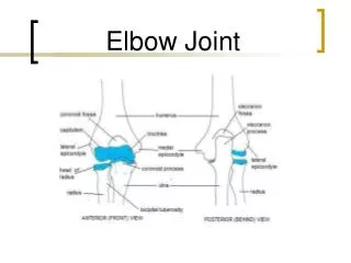
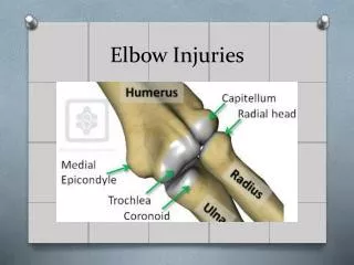
Functional Anatomy • The humerus, radius, and ulna are the 3 bones that make up the elbow. Each bone is designed to allow the elbow to act as a hinge joint. The distal humerus contains the trochlea, capitulum, coronoid fossa, olecranon fossa, radial fossa, medial epicondyle, and lateral epicondyle. The proximal radius includes the radial head, radial neck, and radial tuberosity. The proximal ulna comprises the olecranon process, coronoid process, radial notch, and trochlear notch. • Within the elbow are the ulnohumeral, radiocapitellar, and radioulnar joints, all of which lie within the same joint capsule. The capsule has an internal synovial layer and a superficial fibrous layer. Within these layers are 3 fat pads. The coronoid fossa and radial fossa both contain an anterior fat pad, and the olecranon fossa has a posterior fat pad.
Functional Anatomy The largest of the elbow joints is the ulnohumeral articulation, which is a modified hinge joint. The trochlear groove of the humerus holds the ulnohumeral articulation, which allows for movement between the ulna and the humerus. The radiocapitellar joint is a ball and socket joint composed of the radial head and humeral capitulum. This joint is lateral to the ulnohumeral joint and allows for forearm supination and pronation. The radioulnar joint is a pivot type of synovial joint (a freely movable joint that contains fibrocartilage and hyaline cartilage layers and synovial fluid) divided into superior and inferior sections. The superior section contains the articulation between the radial head and the radial notch of the ulna; the joint rotates within the annular ligament during pronation or supination. The inferior section articulates with the ulnar notch of the radius and swivels around the head of the ulna during pronation or supination.
Functional Anatomy The olecranon process is the bony prominence of the ulna and also is where the triceps muscle attaches to the elbow joint. The olecranon bursa is a fluid-filled sac that provides a cushion between the bone and the slack skin directly over the olecranon process. The bicipitoradial bursa, or cubital bursa, lies between the radial tuberosity and the biceps tendon. The interosseous medial bursa lies medially between the bicipitoradial bursa and the interosseous membrane of the forearm. Below the neck of the radius, the radial tuberosity lies at the insertion point of the biceps tendon. The brachialis muscle attaches at the coronoid process, whereas the radial notch articulates with the radial head to provide radial head stabilization.
Elbow Ligaments • Ligaments provide stability for the elbow joint. The medial and lateral collateral ligaments supply most of the stabilization. The medial collateral ligament (MCL) attaches the ulna to the medial epicondyle of the humerus. The annular ligament loops around the radial head. The lateral ligament attaches the lateral epicondyle to the annular ligament. • The MCL also is called the ulnar collateral ligament (UCL); it has 3 separate bundles and is essential to stabilizing the ulnohumeral articulation of the elbow. The ligament bundles are the anterior, transverse, and posterior bundles. The most important of the elbow stabilizing ligament bundles is the anterior bundle.
Elbow Ligaments The lateral collateral ligament has 4 separate structures: the annular ligament, the accessory lateral collateral ligament, the lateral ulnar collateral ligament, and the radial collateral ligament. The annular ligament encompasses the radial head and stabilizes the radial notch of the ulna by banding the proximal radius to the proximal ulna. The accessory lateral collateral ligament derives from the inferior portion of the annular ligament and connects to the supinator crest of the ulna. The lateral ulnar collateral ligament begins at the lateral epicondyle and also attaches to the ulna’s supinator crest. The radial collateral ligament begins at the lateral epicondyle and inserts into the annular ligament.
Elbow Muscles • Four muscle groups, as well as the tendons, work together to move the elbow joint. The muscle groups are flexors, extensors, the extensor supinator group, and the flexor pronator group. Three muscles within the flexor group primarily act upon the elbow: the biceps brachii, the brachioradialis, and the brachialis muscles. The brachialis muscle and the biceps brachii are the most powerful elbow flexor muscles. • The extensor group contains the triceps and anconeus muscles. The triceps muscle and part of the anconeus muscle control forearm extension. The extensor supinator group contains the brachioradialis, supinator, extensor digitorum, extensor carpi radialislongus and brevis, extensor carpi ulnaris, and extensor digitiminimi. The flexor pronator group includes the pronator teres, flexor carpi radialis, palmarislongus, flexor carpi ulnaris, and flexor digitorumsuperficialis.
Blood Supply and Elbow Nerves • The blood supply through the elbow is extensive, and the major arteries involved with the elbow are the brachial artery, radial artery, and ulnar artery. The brachial artery is lateral to the median nerve and lies within the cubital fossa of the elbow. Within the cubital fossa, the brachial artery then bifurcates into the radial and ulnar arteries. • Four nerves control elbow function and sensation. They are the musculocutaneous nerve, median nerve, ulnar nerve, and radial nerve. The brachial plexus of the elbow is very complex, filled with a network of peripheral nerves. The most accessible nerve is the ulnar nerve. This nerve sits along the olecranon groove and crosses the elbow through the cubital tunnel of the humerus. Because of its location, the ulnar nerve is the elbow nerve injured most often. It most commonly is compressed during direct trauma.
Blood Supply and Elbow Nerves The radial nerve crosses the elbow forward of the lateral epicondyle. Radial nerve compression also is possible because the nerve is susceptible to tightening of the fibrous band that surrounds it. The median nerve runs medially to the biceps tendon and crosses the elbow from within the antecubital fossa. At the elbow, the posterior interosseous nerve branches off of the radial nerve. The musculocutaneous nerve traverses the elbow through the lateral antecubital fossa.
Bone Development • Bones develop in 3 distinct stages: during childhood, adolescence, and young adulthood. • When viewing a radiograph of a pediatric patient’s elbow, it is important to understand the ossification centers or the order in which the elbow joint and bones begin to develop. The order of bone growth is the same for all pediatric patients, and the commonly accepted mnemonic for this order is CRITOE (capitulum, radial head, internal [medial] epicondyle, trochlea, olecranon process, and external [lateral] epicondyle). Ossification centers typically appear in girls 1 to 2 years before they appear in boys.
Radiographic Anatomy • Radiographically, several important anatomical lines are essential in assessing possible elbow damage in patients. The radiocapitellar line should be centered through the long axis of the radius and extend through the radial neck to the center of the capitulum. • A lateral radiograph also should display the anterior humeral line. This line should begin at the anterior portion of the humerus and extend vertically through the middle third of the capitulum. The coronoid line also can be seen on the lateral image. This line should proceed from the top of the coronoid process of the ulna and proximally intersect the anterior portion of the capitulum and trochlea.
Radiographic Anatomy The lateral projection of the elbow also is essential in evaluating the fat pads. The distal humerus contains 2 fat pads that make contact with the joint capsule on the anterior and posterior portion of the joint. Radiographically, this fat pad appears as a darker density next to the bone with a grayer density of tissue surrounding the edges. If the posterior fat pad is noted by the radiologist, this is known as a positive fat pad sign and indicates a probable fracture in approximately 90% of fracture cases. Fat pads appear in the presence of a joint effusion when the capsule that holds the fat pads distends. Correctly positioning the patient’s elbow at 90°is imperative when imaging fat pads. A minor extension of the arm can increase the pressure on the posterior fat pad, making the fat pad appear on the image and leading to a false-positive diagnosis.
Diagnostic Imaging Modalities: Radiography • The most common findings are fractures, arthritis, loose bodies, and destructive processes. Radiologic technologists should obtain a minimum of 2 projections with 90°of differentiation because the elbow anatomy appears normal in some projections even when the patient has a disorder or injury in the area. • For the elbow, an anteroposterior (AP) projection and a lateral projection should be taken. Other common elbow projections include the medial and lateral oblique, radiocapitellar, and the Jones method, also known as the distal humerus acute flexion projection.
Radiography In the AP projection, the patient’s elbow is extended over the cassette. Supination of the hand prevents the forearm bones from crossing. A slight lateral tilt of the forearm can place the anatomy in the correct position. The x-ray beam is perpendicular and centered to the elbow joint. When the patient cannot completely straighten the elbow, 2 images replace the AP projection. Positioning for both projections is similar to the AP projection, in that the central ray is perpendicular to the joint. However, the first projection places the posterior surface of the humerus flat and parallel to the cassette. For the second projection, the technologist places the patient’s arm so that the posterior forearm is flat and parallel to the cassette.
Radiography Lateral images of the elbow require that the patient flex the elbow 90°. Both the forearm and the humerus should be parallel to the surface of the cassette during contact, and the radiologic technologist should rotate the patient’s hand into a true lateral position. The central ray should be directed perpendicular to the elbow joint. The medial oblique projection requires the patient to extend the elbow over the cassette as in the AP projection but with the arm and hand in a pronated position. The internal oblique projection is positioned similarly, but the arm should be rotated laterally until the elbow is at a 45°angle to the cassette.
Radiography The radiocapitellar projection requires that the forearm be imaged using a lateral projection but that the hand and arm remain in a neutral rotation. The x-ray beam points at a 45°cephalic angle toward the shoulder, centering on the elbow joint. The radiocapitellar projection shows an oblique angle of the lateral elbow separating the proximal radius and ulna. The Jones method acquires images of the elbow in complete flexion. The posterior aspect of the humerus lies on the cassette with the forearm superimposed over the top. The x-ray beam should be perpendicular to the cassette and centered approximately 2 inches above the olecranon process.
Magnetic Resonance Imaging • Magnetic resonance (MR) imaging helps display the joint’s muscle and tendon attachments. The ability to evaluate much of the elbow anatomy would make MR an optimal imaging choice except that positioning the elbow is difficult using MR imaging. • For elbow MR, the patient can lie on his or her stomach with the arm positioned above the head. Patients do not tolerate this position well; therefore, the supine position is used more frequently. • The classic MR acquisition for the elbow involves axial, coronal, and sagittal images with T1- and T2-weighted images.
Magnetic Resonance Imaging MR images of the 3 major nerves of the elbow generally appear the same intensity as muscle on T1-weighted imaging. The signal intensity is slightly higher with T2-weighted images, but outlining and visualizing the nerves depend somewhat on adjacent fat. MR images of an injured ligament might show thickening or thinning of the ligament, increased signal intensity, hemorrhage, slackness, and other abnormalities. Muscle injuries on MR scans show morphological changes, atrophy, fatty changes, and edema. Joint fluids increase with diseases that produce synovial inflammatory changes. MR using gadolinium contrast enhances the tissue’s signal intensity.
Computed Tomography • The rapid scanning and helical imaging of modern CT scanners make accurate and prompt imaging of elbow trauma possible. Planning for elbow surgery also benefits from CT’s ability to reformat images in any plane required and to provide 3-D volume renderings. • CT displays fractures, loose bodies, osteochondral lesions, and other bony abnormalities well. Aside from fracture fragment evaluation, CT with IV contrast also is beneficial for blood vessel evaluation following trauma. Similar to conventional arthrography of the elbow, CT arthrography can highlight the joint capsule and filling defects from synovitis or loose bodies. CT arthrography also is helpful in evaluating MCL tears.
Ultrasonography • Ultrasonography is not a typical choice for imaging the elbow, but the modality offers a less expensive alternative for evaluating tendons, ligaments, and nerves. Ultrasonography is useful in diagnosing soft-tissue diseases of the elbow. It is also a good choice when imaging infants and young patients to evaluate unossified epiphyses that might not be noticeable on radiographs. • Color-flow Doppler imaging can highlight soft-tissue inflammation by showing increased blood flow to areas of the elbow. Doppler imaging also can help distinguish cystic from solid masses by displaying the vascular components of the mass. • Ultrasonography is useful for guiding aspiration needles or during therapeutic measures for the elbow.
Elbow Disorders: Olecranon Bursitis • Inflammation of the olecranon bursa is called olecranon bursitis. It is also referred to as student’s elbow because the condition can be caused by leaning excessively on the elbow. Chronic olecranon bursitis is seen in people who throw repetitively, such as baseball pitchers; acute cases usually occur after a direct fall onto a hard surface. • Patients with bursitis are easily identified by the large amount of swelling and masslike appearance of the elbow. In nontraumatic situations, imaging may not be required if the bursa fluid can be aspirated. In traumatic situations, however, the bursa can become inflamed because of an olecranon fracture. Fluid aspiration sometimes leads to infection, and continued pain should be investigated with further imaging studies.
Cubital Bursitis • Cubital bursitis also is known as bicipitoradial bursitis, and symptoms include antecubital fossa swelling and tenderness. • MR imaging of cubital bursitis shows high-signal fluid that emerges between the radial tuberosity and biceps tendon distally. The fluid normally appears on T2-weighted images, which can lead to misdiagnosis of a soft-tissue tumor. In questionable cases, IV contrast assists in the diagnosis. If the fluid collection does not enhance, bursitis is the likely diagnosis. • During sonography, elbow extension is routine. Sonograms of cubital bursitis can show fluid or hypoechoic tissue that causes active distention of the bicipitoradial bursa. Doppler imaging shows an increase in blood flow to the area, which is symptomatic of active inflammation.
Tendonitis and Tendon Tears • Tendonitis of the elbow is due to inflammation of the tendons. Tendonitis of the biceps muscle can lead to rupture on either end. This condition commonly occurs after lifting heavy objects and results in tenderness near the biceps rupture site. On average, tendonitis of the biceps occurs in men aged 45 to 60 years. • When the distal end of the biceps muscle ruptures, symptoms include proximal elbow pain and weakness, especially during supination. When the rupture occurs, the patient can experience a snapping sensation followed by the appearance of a bulbous deformity, or “Popeye sign,” near the distal bicep. • Radiographs may show an avulsion fracture of the radial tuberosity in cases of complete tears, but enlargement or abnormality of the radial tuberosity is the most common finding. MR imaging is useful to assess a possible tear or degeneration of the biceps tendon. Ultrasonography may be helpful in determining the extent of any tears.
Tendonitis and Tendon Tears Triceps tendonitis is common with repetitive elbow use in young athletes. The individual often experiences a sensation on the medial border of the elbow that patients describe as something snapping into place. As with bicep tears, MR and ultrasonography images can help physicians distinguish tears from other pathology.
Lateral Epicondylitis • Lateral epicondylitis is the most common sports-related injury of the elbow and a primary cause of elbow pain. The mechanism of injury depends on repeated, forceful contraction of the wrist extensor muscles; contraction occurs with frequent forearm pronation and supination, along with wrist extension. The abuse of the extensor muscles causes inflammation at the lateral epicondyle. • Lateral epicondylitis is frequently referred to as tennis elbow. Although the condition is associated with tennis, many other repetitive motions can cause epicondylitis. Estimates show that 90% of lateral epicondylitis patients develop the disorder from activities other than tennis.
Lateral Epicondylitis The pain felt with lateral epicondylitis normally occurs at the lateral epicondyle or slightly outside the elbow. Gripping items may become difficult because of weakness and increased pain within the forearm. This is known as the “coffee cup” sign. Radiographic evidence of lateral epicondylitis is rarely found. In any case, radiographs differentiate lateral epicondylitis from other disease processes of the elbow such as arthritis or loose bodies.
Lateral Epicondylitis Regardless of whether the tendonosis is located medially or laterally, MR imaging demonstrates the same epicondylitis features on either side. Ultrasonography can find calcifications or hypoechoic areas in the lateral epicondyle region and may be useful in diagnosing tendonosis. In cases of chronic lateral epicondylitis, the capsule below the extensor carpi radial brevis (ECRB) tendon should be examined for tears. Currently, the best modality for diagnosing capsular tears is arthroscopic techniques. Although MR falls short of accurately imaging capsular tears of the ECRB tendon, CT arthrography has shown excellent success at displaying capsular tears.
Medial Epicondylitis • Known to patients as golfer’s elbow, medial epicondylitis is common in individuals who overuse their wrist flexors and forearm pronator but is seen far less frequently than lateral epicondylitis. • Medial epicondylitis primarily affects the insertion point of the flexor carpi radialis. The patient presents with pain at the medial aspect of the elbow. • As with lateral epicondylitis, radiographic evidence of medial epicondylitis can be difficult to find, but small calcifications or spurs next to the medial epicondyle are common. MR imaging most often is used for diagnosis.
Arthritis • Arthritis is a joint disorder that results in inflammation of the joints within the body. There are varying kinds of arthritis, each exhibiting different causes and symptoms. Regardless of the type of elbow arthritis, the initial stages of pain management include pain medication and physical therapy. Occasionally, corticosteroid injections are used for pain management, but physicians must use caution to ensure that the injections do not lead patients into a false sense of treatment and overuse of the elbow joint, causing further deterioration.
Rheumatoid Arthritis • Rheumatoid arthritis is a severe form of arthritis that progressively affects the body’s joint tissues. Joint erosion and destruction are common because of the severity of rheumatoid arthritis. • Rheumatoid arthritis commonly begins in the radiocapitellar joint. The radial head may move out of its regular position and cause problems with other elbow anatomy. • Radiographs can monitor structural changes caused by rheumatoid arthritis, but radiography is not the preferred method for early disease assessment. Bone erosion from rheumatoid arthritis is better displayed on CT images than on MR images or radiographs. Ultrasonography can show inflammation related to rheumatoid arthritis and document destructive changes.
Osteoarthritis • Arthritic conditions of the elbow are not uncommon, but osteoarthritis of the elbow is rare. Osteoarthritis is much more prevalent in weight-bearing joints, such as knees and hips, and in the interphalangeal joints of the hand. • Patients with osteoarthritis affecting the elbow experience the most pain during terminal flexion and extension of the joint. • Radiographic evidence of osteoarthritis includes osteophyte formation. These osteophytes usually are near the ulnohumeral joint and occasionally impinge on the ulnar nerve. MR and CT images of the elbow help show the joint surfaces and detect loose bodies or spurs.
Gout • Elevated uric acid levels can result in monosodium urate crystals to infiltrate the synovial fluid of joint spaces and lead to gout. Gout usually is found in the joint spaces of the toes but can appear at the elbow. • Evidence of gout is obvious in patients with advanced disease but is not often apparent on images of early cases. MR images are better for evaluating synovial involvement, and CT is better for displaying intraosseous lesions. Ultrasonography also can highlight thickening of the synovial fluid, along with inflammation.
Overuse Conditions in Children • Children also can have elbow injuries and conditions related to overuse of the elbow joint. Examples of these problems, sometimes referred to as Little League elbow, include traction apophysitis of the medial epicondyle, Panner disease, and osteochondritisdissecans.
Traction Apophysitis of the Medial Epicondyle • Traction apophysitis of the medial epicondyle is inflammation of the medial epicondyle due to an avulsion tear or trauma. Because of the timing of ossification in children, traction apophysitis is the most common elbow injury in young children. • Approximately 97% of elbow problems in baseball pitchers are associated with symptoms of the medial elbow. The main symptom of apophysitis of the medial epicondyle is pain immediately after a repetitive motion such as throwing. • Radiographs of the elbow might show slight widening of the apophysis, but this can be missed easily if the radiologist does not review comparison radiographs of the opposite elbow. MR imaging can show edema of the bone marrow around the medial epicondylar area.
Panner Disease • Osteochondrosis affects the ossification centers of children when the bone degenerates and then begins to regenerate, producing excessive calcification in some areas. Panner disease is a form of osteochondrosis that affects the capitulum of the elbow. • Panner disease is most common in preadolescent boys and children younger than 10. Panner disease is the most common reason for lateral elbow pain in young children and routinely affects the dominant arm. • Radiographs can display sclerosis and areas of decreased density at the capitulum. An MR series with T1-weighted images might show fragmentation with decreased signal intensity at the capitulum surface.
Osteochondritis Dissecans • A more advanced form of osteochondrosis is osteochondritisdissecans. These lesions result from death of the articular cartilage or subchondral bone of the capitulum due to a lack of blood supply. Subchondral bone provides cartilage support at articulation sites. If the subchondral bone or cartilage is slightly fractured, it is known as osteochondritisdissecans. • Patients with these osteochondritisdissecans lesions often have plica, an inflamed lining of the radiocapitellar joint. Because the pain is located on the lateral side of the elbow, radiocapitellarplica commonly is misdiagnosed as lateral epicondylitis.
Osteochondritis Dissecans • Osteochondritis dissecans typically affects adolescent athletes aged 11 to 21 years. • Osteochondritis dissecans is characterized by a dull ache with no centralized location. The pain may disappear when the child is resting then reappear during strenuous activities. If fragments are loose in the area, the joint may routinely catch or cause a popping sensation.
Osteochondritis Dissecans Radiographs typically are not very sensitive for identifying loose bodies, and less than 30% of positive loose bodies are found using radiography. CT of the elbow joint can help physicians locate and count loose bodies found throughout the elbow compartments before the patient undergoes arthroscopic procedures to remove the fragments. On MR images, osteochondral lesions can be confused with normal osseous variants of the elbow. Ultrasonographic imaging of osteochondritisdissecans is rare. Arthroscopy is used most often because it remains a safe and effective way to diagnose and evaluate osteochondritisdissecans or lesions of the elbow. The benefit of arthroscopy is the ability to immediately remove any loose bodies or deliver other treatments.
Nerve Damage • The most common nerve condition in the elbow is ulnar neuritis. This inflammation of the nerve can cause radiating pain from the posterior medial elbow to the hand and fingers. Compression of the elbow nerves is attributed to a wide range of abnormalities. • An ulnar nerve injury typically is associated with supracondylar fractures when the arm is in hyperflexion, but most ulnar nerve injuries occur because of direct blows to the nerve.Damage to the ulnar nerve can lead to numbness and tingling of the hand. • In severe cases of ulnar nerve injury, symptoms can last for years after the trauma. This is commonly known as tardy ulnar palsy. • MR imaging is the best modality to show nerve damage and edema.
Elbow Injuries: Ligament or Tendon Injury • About 50% of the medial and lateral plane of the elbow is stabilized by ligaments. If the anterior bundle of the elbow’s MCL is injured, the elbow becomes extremely unstable except when fully extended. • Radiography can assess ligament tears and joint stability, particularly with valgus or varus stress applied during a fluoroscopic examination. • Gadolinium contrast often is used during MR imaging to help enhance MCL injuries. Although MR imaging is highly sensitive for complete tears of the MCL, it is not the method of choice for viewing partial tears. CT arthrography has a higher rate of identifying partial tears. In either modality, a sagittal projection is the best choice for determining the size of a ligament injury.
Dislocations • The elbow is the joint that is dislocated most often among pediatric patients. It is the second most dislocated joint in adult patients, with 50% of cases resulting from sports activities. • Elbow dislocations can occur as a result of many situations but most often arise from a fall on an outstretched hand. The most common dislocation involves displacement of both the ulna and radius. • Elbow dislocations often present with other injuries. The MCL is routinely compromised in elbow dislocation. With MCL damage, the elbow joint might remain unstable unless the ligament heals or is surgically repaired. • A patient with an elbow dislocation most often experiences extreme pain and swelling. A deformity may or may not be clearly seen.
Dislocations An AP and lateral radiograph elbow series is sufficient to diagnose an elbow dislocation. An elbow reduction most often is completed by having the patient’s upper arm held in place while applying a steady pull on the forearm and hand. Once the dislocation is reduced, the elbow should be tested for mobility and examined with radiography to ensure satisfactory reduction of the joint and exclude any further bone injury. A neurovascular examination should be performed and results noted before and after reducing the elbow joint dislocation. Following an elbow dislocation, extension may become compromised and the elbow may be unstable. Over time, some patients might develop arthritis or ectopic bone.
Heterotopic Bone Formation • Formation of bone or calcification that is not in the normal bone growth area is called heterotopic bone formation. Heterotopic bone growth at the elbow can be associated with traumatic injury of the elbow, but heterotopic bone formation of the elbow also can be caused by central nervous system trauma or excessive burns. • The formation of juxta-articular bone at the elbow joint can cause problems because of decreased full range of motion. Once elbow motion has been compromised by heterotopic bone formation, the only treatment for restoring motion is surgical resection of the bone.
Fractures • The proximity of nerves, arteries, tendons, muscle, and bones in the elbow contributes to the elbow being considered one of the most complex fracture sites. Clinical examination begins by observing the patient’s arm. When both arms hang normally at the patient’s sides, there should be a 5° to 15°separation of the forearms and hands from the body. This arm-to-body separation is known as “the carrying angle.” If the patient’s arms and hands are not observed within the acceptable ranges, it could indicate an elbow fracture. Any variation of the angle that is more than 15°is known as cubitus valgus. Angles less than 5°are called cubitusvarus. • All fractures are serious and should be treated as such, although open fractures are at higher risk for adverse complications. • There are three distinct phases in the healing process of bones and complete fracture healing can last for months or years.
Distal Humerus Fractures • Distal humerus fractures represent only 2% of adult fractures. These fractures can appear in both condyles and often continue into the joint space. • Location determines the classification of distal humerus fractures. • Patients with distal humerus fractures experience extensive swelling, deformities, and pain. These fractures also can produce bruising of the skin. Flexion of the arm may produce crackling or popping sounds from bone fragments.
Distal Humerus Fractures • A 2-projection radiographic examination usually is sufficient to evaluate distal humerus fractures. A lateral oblique projection is helpful for diagnosing lateral condyle fractures or displacement. An oblique radiograph of the elbow might help identify nondisplaced fractures of the condyle in children. • Posterior displacement of the humerus, often seen in fractures, affects the orientation of the anterior humeral line seen on the lateral image. If the anterior humeral line passes through the anterior portion of the capitulum or does not meet the capitulum at all, a fracture is possible. • Displaced fractures are generally stabilized with surgery. Splinting is sufficient in nondisplaced fractures. It is important to retake radiographs of the elbow after several days of splinting to ensure adequate positioning.