Ventilators
Ventilators. Kindred Hospital Louisville Education Module. Learning Objectives. Identify the mechanics of breathing Identify indicators for mechanical ventilation Identify two types of ventilators Identify the Modes of Ventilation Discuss the Adjuncts to Mechanical Ventilation

Ventilators
E N D
Presentation Transcript
Ventilators Kindred Hospital Louisville Education Module
Learning Objectives • Identify the mechanics of breathing • Identify indicators for mechanical ventilation • Identify two types of ventilators • Identify the Modes of Ventilation • Discuss the Adjuncts to Mechanical Ventilation • Identify the components of Ventilator Settings • Describe the Nursing Care of the Mechanically Ventilated Patient • Discuss Arterial Blood Gases
Components of Respiratory System • Nasal & Oral Cavities • Nasopharynx • Oropharynx • Epiglottis • Larynx • Trachea • Left & Right Bronchus • Left & Right Lung • Alveoli
Pathophysiology of Breathing • During breathing, air is inhaled through the airway into millions of tiny sacs where gas exchange takes place (alveoli). Then the air mixes with the carbon dioxide-rich gas coming from the blood. This air is then exhaled back through the same airways to the atmosphere. Normally this pattern repeats itself from 12 - 20 times a minute, but can increase or decrease to meet our body’s needs. • The gas exchange that takes place as described above is the main function of the lungs. It is required to supply oxygen to the blood for distribution to the cells of the body, and to remove the carbon dioxide that the blood has collected from the cells of the body.
Pathophysiology of Breathing • Gas exchange in the lungs occurs only in the smallest airways and the alveoli. It does not take place in the conducting airways (pathways) that carry the gas from the atmosphere. The volume of these conducting airways is called the anatomical “dead space” because it does not participate directly in the gas exchange. • Gas is carried through the conducting airways through a process called “convection”. • Gas is exchanged between the alveoli and the blood through “diffusion”. • In normal, healthy lungs the drive to breathe comes from the need to regulate carbon dioxide levels in the blood, not from a desire to inhale oxygen.
Pathophysiology Cont’d • One of the biggest factors that determines whether breathing is producing enough gas exchange to keep a person adequately oxygenated is the ‘ventilation’ that each breath is producing. • Ventilation is expressed as the volume of gas entering or leaving the lungs in a given amount of time. It can be calculated by multiplying the inhaled (or exhaled) volume of a gas (Tidal Volume) times the breathing rate. • For example: A person breathing in 0.5 Liters per respiration, who breathes 12 times a minute, has a volume of 6 Liters/minute
Pathophysiology Cont’d • During normal breathing, the body selects a combination of tidal volume that is large enough to clear the dead space and add fresh gas to the alveoli, and a breathing rate that ensures the correct amount of ventilation is produced. • There are two sets of forces that can cause the lungs and chest wall to expand: the forces that are produced by the muscles of respiration when they contract; and the force produced by the difference between the pressure at the airway opening and the pressure on the outer surface of the chest wall. • In normal respiration, the muscular force is the only one that comes into play, when the respiratory muscles do the needed work to expand the chest wall, decreasing the pressure on the outside of the lungs so they expand, which draws air into the lungs.
Pathophysiology Cont’d. • When respiratory muscles are not able to do the work required for ventilation, the pressure at the airway opening, and/or the pressure at the outer surface of the chest wall can be manipulated to produce breathing movements. • When altering either of those pressures, you can do so in one of two ways. Either increase the pressure at the mouth and nose, so that air is forced into the lungs; or lower the pressure on the chest wall external surface.
Breathing Pathophysiology • Remember that an alteration in any of the areas associated with breathing/gas exchange can produce undesirable effects in your patient’s oxygenation. • Within the chest wall, there is normally a constant negative pressure that facilitates respiration. If this negative pressure is disrupted, ventilation and oxygenation are disrupted.
Indications for Mechanical Ventilation • Acute dyspnea • Significant respiratory acidosis • Acute or impending ventilator failure (elevated PaCO2 {>50 mmHg} with a pH < 7.30) • Severe oxygenation deficit despite high supplemental oxygen delivery (PaO2 < 60 mmHg on FiO2 > 60%) • Secretion/Airway Control • Apnea, Respiratory Arrest
Common Diseases Requiring Mechanical Ventilation • Acute Obstructive Disease: acute severe asthma; airway mucosal edema) • Altered Ventilatory Drive: hypothyroidism; intracranial hemorrhage; dyspnea-related anxiety • Cardiopulmonary Problems: CHF; Pulmonary Hemorrhage • Chronic Obstructive Pulmonary Disease: emphysema, chronic bronchitis, asthma; cystic fibrosis; bronchiectasis • Neuromuscular Disease: ALS; Guillian-Barre; Cancer; Malnutrition; Infections • Atelectatic Disease: ARDS; Pneumonia
Other Common Conditions Requiring Mechanical Ventilation • Burns and Smoke Inhalation: inhalation injury, surface burns • Chest Trauma: Blunt injury; flail chest; Penetrating Injuries • Fatigue/Atrophy: Muscle overuse; disuse • Head/Spinal Cord Injury: Meduallary brainstem injury; Cheyne-Stokes breathing; Neurogenic Pulmonary Edema • Postoperative Conditions: Cardiac & Thoracic Surgeries; • Pharmacological Agents/Drug Overdose: Muscle relaxants; barbiturates; Ca+ channel blockers; long-term adrenocorticosteroids; aminoglycoside antibiotics
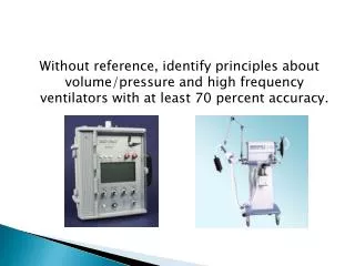
POSITIVE PRESSURE VENTILATION Uses the technique of applying positive pressure (relative to atmospheric pressure) to the airway opening NEGATIVE PRESSURE VENTILATION Uses the technique of applying negative pressure (relative to atmospheric pressure) to the external body surface Two Approaches to Mechanical Ventilation
Positive Pressure Ventilators Simplified • For safe operation of the ventilator, the following things are required: • Patient Interface: The ventilator delivers gas to the patient through a set of flexible tubes called a patient circuit. This can have one or two tubes. The circuit typically connects the ventilator to the patient to either an endotracheal tube or tracheostomy tube. • Power Sources: Typically these are powered by electricity or compressed gas. The ventilator is usually connected to separate sources of compressed air and compressed oxygen. Because compressed gas has all the moisture removed, a humidifier is needed to moisten the gases being delivered to the patient. • Control System: This ensures the patient receives the desired breathing pattern. It involves setting the parameters of the size of the breath, how fast it is brought in & out, and how much effort the patient must exert to signal the ventilator to start a breath. • Monitors: A pressure monitor, as well as volume and flow sensors to provide alarms if readings are outside the desired range.
Negative Pressure Ventilators Simplified • For safe operation of the automatic ventilator, the following things are required: • Patient Interface: The patient is placed inside a chamber with his or her head extending outside the chamber. The chamber may encase the entire body except the head(iron lung) or it may enclose just the rib cage and abdomen (cuirass: pronounced cure-ahs). It is sealed to the body where the body where the body extends outside the chamber. • Power Sources: Electricity powered, to run a vacuum pump that periodically evacuates the chamber to produce the required negative pressure. • Control System: Sets breathing patterns. • Monitors: Alarms.
Modes of Mechanical Ventilation • Controlled Mandatory Ventilation: (CMV) The patient receives a set respiratory rate at set time intervals with a consistent tidal volume. This is generally only used with much sedation or paralytics, because patient efforts do not trigger the delivery of a breath by the machine. This is used when the patient must not expend energy to breathe.
Modes of Mechanical Ventilation • Assist Control: (AC) The patient receives a set respiratory rate at set time intervals with a consistent tidal volume, but when the patient initiates a breath on their own, the preset tidal volume is delivered. This decreases the patient’s effort of breathing, and ensures volume delivery.
Modes of Mechanical Ventilation • Synchronized Intermittent Mandatory Ventilation (SIMV): The patient receives a preset respiratory rate at a set tidal volume, but the machine allows for the patient to breathe spontaneously during the machine breaths. If the patient breathes near the time that the machine is prepared to deliver the preset volume, the machine will deliver the preset tidal volume. The breaths that the patient initiates in between the machine breaths are not supplemented by the machine. It is usually tolerated well by the patient, because of the synchronicity involved.
Modes of Mechanical Ventilation • Continuous Positive Airway Pressure (CPAP): Used either intermittently during long-term weaning as a way to strengthen the muscles, or as a final step before removing the patient from the ventilator, to see how they tolerate the lack of ventilatory assistance. All breaths are generated by the patient, and the patient’s effort determines the tidal volume. The machine simply provides a continuous airway pressure, supplemental oxygen, and apnea alarms. The continuous airway pressure makes the effort of breathing easier for the patient.
Modes of Ventilation • Pressure Support (PS): When this mode is used, the patient initiates the breath, and the inspiration ends when a preset flow amount is delivered. The positive pressure is applied throughout inspiration and helps to increase the amount of tidal volume the patient “pulls in” and decreases the energy the patient has to use.
Adjuncts to Mechanical Ventilation • Positive End Expiratory Pressure (PEEP) • PEEP is the application of continuous airway pressure throughout expiration. The presence of this pressure in the airway prevents the complete collapse of the alveoli, and helps maintain that pressure until the next inspiration cycle begins.
Components of Ventilator Settings • Rate • Tidal Volume • Percentage Oxygen • Peep or Pressure Support
Rate • The rate is the number of times the ventilator is set to provide a breath to the patient. This may vary from 8-20 breaths per minute.
Tidal Volume • Tidal volume is the amount of gas the the ventilator is to provide to the patient with each breath. This volume will vary based on each patient’s height, weight, and gender. To calculate a very rough estimate of tidal volume, you can use 10 - 15cc per kilogram of body weight. So a 75lb. patient might have an ordered tidal volume of 750cc.
Percentage of Oxygen • The percentage of oxygen supplied to the patient with every breath. This can be as low as 40% to as much as 100%. Higher oxygen percentages for long periods of time increase the patient’s risk for oxygen toxicity and other pulmonary complications.
PEEP • PEEP can be added to the regular ventilator settings, to provide the positive end expiratory pressure that helps to prevent the completecollapse of the alveoli.
Pressure Support • The patient initiates the breath, and the inspiration ends when the preset flow target is delivered. The tidal volume will vary, depending on the patient. The positive pressure is applied throughout inspiration and helps the patient to “pull in” the tidal volume, and reduces their energy expenditure.
Nursing Care of the Mechanically Ventilated Patient • Nursing care of patients who are being mechanically ventilated requires some special considerations. • Some special considerations relate specifically to the type of tube via which the patient is being ventilated (i.e. endotracheal or tracheostomy) and others related to the patient, and the ventilator itself.
Nursing Care of the Patient with an Tracheostomy Tube • Trach care should be performed at least every shift, and as needed as ordered by the patient’s Physician. • The patient should always be pre-oxygenated with 100% oxygen prior to suctioning. • Saline should not be routinely instilled into the airway. Saline installation has been shown to increase infection rates and to cause decreased oxygen levels for longer periods of time than suctioning without it.
Nursing Care of the Mechanically Ventilated Patient • Pulmonary assessment is perhaps never as important as it is in the mechanically ventilated patient. • These patients require frequent reassessments on a schedule and on an “as needed” basis. • Further assessments can be documented in Protouch under “Reassessments”.
Nursing Assessment Components: Breath Sounds • Breath sounds should be assessed at least every four hours, and more frequently as needed. • Both the anterior and the posterior chest need to be auscultated bilaterally. • Clearly document any adventitious breath sounds that are heard, and report significant alterations to the Physician.
Nursing Assessment Components: Rate & Volume • Make sure to assess and document the patient’s spontaneous respiratory rate and tidal volume. This information tells you a lot about the patient’s respiratory functioning. • Note any changes in this area, and report significant findings to the patient’s Physician.
Nursing Assessment Components:Pulse Oximetry • Pulse oximetry is a useful monitoring tool, but provides minimal indication of the patient’s ventilatory or acid-base status. • Readings can be affected by abnormal hemoglobins, vascular dyes, and poor perfusion. • Plus, the machine can’t distinguish between normal and abnormal hemoglobins, so a patient with carbon monoxide poisoning could have a pulse ox reading of 100%.
Nursing Assessment Components:Sputum • A respiratory system assessment should include documentation of any sputum. • Note the color; tenacity; odor; frequency; quantity; of sputum for a thorough assessment. • Note if the patient is able to expectorate his/her own sputum, or if suctioning is required to remove it.
Complications of Mechanical Ventilation • One of the reasons for such a frequent and thorough assessment of the pulmonary system while patients are being mechanically ventilated is due to the many complications that can occur with the use of mechanical ventilation. • Thorough assessments can lead to the early discovery of potential complications, heading off more serious complications later.
Complications of Mechanical Ventilation • Positive Pressure Ventilation: • can cause:hypotensiondecreased venous returndecreased cardiac output • Other complications:pneumothoraxsubcutaneous emphysemaair emboluslocalized pulmonary hyperinflationnosocomial infectionsincreased intracranial pressure (cerebral edema)
Understanding ABG’s are critical to understanding the respiratory status of the patient. As a nurse, it is essential you have a working knowledge of ABG’s. That responsibility cannot be “delegated” to R.T. ABG Components: pH PCO2 HCO3 Base Excess/Deficit PaO2 ABG Overview
pH • pH is the relative acidity or baseness of the blood. • Normal human blood pH ranges from 7.35 - 7.45 • Less than 7.35 is considered acidotic and greater than 7.45 is considered alkalotic
Conditions that alter the pH of blood fall into one of four processes. One or more of these processes may be present in a patient with an abnormal acid-base status. Four Processes: Metabolic Acidosis Metabolic Alkalosis Respiratory Acidosis Respiratory Alkalosis pH
Metabolic Processes • Metabolic processes are those that primarily alter the bicarbonate concentration in the blood. A decrease in the blood concentration of bicarbonate leads to metabolic acidosis, while an increase in serum bicarbonate levels leads to metabolic alkalosis.
Respiratory Processes • Respiratory processes alter the pH of the blood, by changing the carbon dioxide levels. Carbon dioxide that accumulates in the blood causes an acid state (carbonic acid). • As respirations increase or decrease in rate, the level of carbon dioxide in the blood varies. Faster respirations cause decreased blood carbon dioxide levels, and slower respirations cause less carbon dioxide to be “blown off”, causing an increased serum carbon dioxide level.
Respiratory Processes • Respiratory alkalosis occurs when respirations increase, leaving less carbon dioxide in the blood, and when respirations decrease, the carbon dioxide level in the blood increases, which can lead to respiratory acidosis.
PCO2 • PCO2 is the partial pressure of dissolved carbon dioxide in the blood. • Most is excreted by the lungs, some is excreted in the kidneys as HCO3. • Normal level is 35 - 45 mmHg. • PCO2 level is a direct indicator of the effectiveness of ventilation • As PCO2 rises, the blood becomes more acidic and the pH drops • As PCO2 decreases the blood becomes more alkaline an pH rises • If a change in the PCO2 level is the primary alteration, then a respiratory problem exists
HCO3 • Bicarbonate is the primary buffer in the body. Buffers neutralize acids. • Normal range is 22 - 26 mmHg. • As the HCO3 level rises, the blood becomes more alkaline and the pH increases. • As the HCO3 level falls, the blood becomes more acidic and the pH decreases. • If a change in HCO3 is the primary alteration, then a metabolic problem exists.
Base Excess/Deficit • Measures the excess amount of acid or base present in blood. This is independent of changes in PCO2, so it’s a measure of metabolic acid-base balance. • Increased HCO3 = base excess (alkalosis) • Decreased HCO3 = base deficit (acidosis)
PO2 • The amount of oxygen dissolved inplasma • Normal is 80 - 100 mmHg in healthy people breathing room air at sea level. • Normal PO2 will decrease with altitude and aging. • PO2 > 60mmHg may be considered acceptable in critically ill, mechanically ventilated adults. • Adequacy of PO2 must be weighed against the potential for oxygen toxicity
Analyzing Blood Gas Results • Use the following simple four step process to interpret ABG’s. • Practice this until you are completely comfortable with it. • Keep a “cheat sheet” with this information written down and refer to it! • Practice, Practice, Practice!!!