Introduction to Mechanical Ventilators
1.65k likes | 9.94k Vues
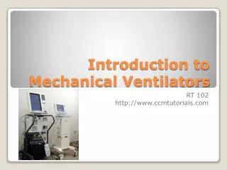
Introduction to Mechanical Ventilators. RT 102 http://www.ccmtutorials.com. A mechanical ventilator is a machine that generates a controlled flow of gas into a patient’s airways.

Introduction to Mechanical Ventilators
E N D
Presentation Transcript
Introduction to Mechanical Ventilators RT 102 http://www.ccmtutorials.com
A mechanical ventilator is a machine that generates a controlled flow of gas into a patient’s airways. • Oxygen and air blended according to the prescribed inspired oxygen tension (FiO2), accumulated in a receptacle within the machine, and delivered to the patient using one of many available modes of ventilation. • The ability to initiate, maintain and wean from the ventilator is the responsibility of the RT • As soon as you initiate the ventilator you must be thinking how to wean the patient off What is a Ventilator
The central premise of positive pressure ventilation is that gas flows along a pressure gradient between the upper airway and the alveoli. • The magnitude, rate and duration of flow are determined by the operator. Flow is either volume targeted and pressure variable, or pressure limited and volume variable. • The pattern of flow may be either sinusoidal (which is normal), decelerating or constant. Flow is controlled by an array of sensors and microprocessors. Conventionally, inspiration is active and expiration is passive (although modern ventilators have active exhalation valves). What is a Ventilator
There are two phases in the respiratory cycle, high lung volume and lower lung volume (inhalation and exhalation). Gas exchange occurs in both phases. Inhalation serves to replenish alveolar gas. • Inspiration increases tidal volume and intrathoracic pressure. Co2 is “washed out” What is a Ventilator
Failure to oxygenate is caused by reduced diffusing capacity and ventilation perfusion mismatch. This can often be overcome by restoring FRC by increasing baseline airway pressure using CPAP. If the problem is atelectasis due, for example, to mucus plugging or diaphragmatic splinting following abdominal surgery, or moderated amounts of pulmonary edema, CPAP, as delivered by facemask or endotracheal tube, may sufficiently restore pulmonary mechanics to avoid addition inspiratory support. • If oxygenation problems are associated with ventilation problems a more aggressive strategy of support is needed
Transairway Pressure: gradient between airway opening and alveoli (Airway pressure – Alveolar pressure). Represents changes in the resistance to gas flow in airways • Transthoracic Pressure: alveolar space and body surface. Represents pressure needed to expand or contract the lungs and chest wall at the same time • Transpulmonary Pressure (Pl): difference between alveolus and pleural pressure/ called alveolar distending pressure. All modes increase Pl pressure by decreasing Ppl Pressure Gradients
Positive pressure changes the normal passive negative pressures found in the lung/pleural space. • During inspiration the pleural space with positive pressure changes from -10 to + 5 • Compliance, elasticity and resistance become very important aspects with the application of positive pressure. • Know Static/Dynamic compliance, RAW Pressure Gradients
Positive Pressure ventilation: moves positive pressure into airway via pressure gradient • High Frequency Ventilation: uses above normal ventilating rates with below normal volumes. 3 modes (HFPPV rates 60-100) (HFJV rates 100-600) (HFOV rates up to 4000) • Negative Pressure Ventilation (iron lung) Types of mechanical ventilation
Support/manipulate pulmonary gas exchange • Alveolar ventilation: normal or permissive hypercapnia • Alveolar oxygenation: maintain CaO2 • Increase Lung volumes: prevent ATX, restore FRC, reduce WOB • Reverse acute respiratory failure/distress/hypoxemia Objectives of mechanical ventilation
Respiratory Failure: Characterized by increased arterial carbon dioxide tension, or failure to oxygenate, characterized by decreased arterial oxygen tension • Impending respiratory failure: Progressing disease, weakening diaphragm, inability to compensate for a severe metabolic acidosis. • Apnea, Post operation sedation • Cardiopulmonary arrest • Spinal cord injury, paralysyis Indications for the ventilator
MIP -20 to 0 cmH2O • MEP Less than 40 cmH2O • VC Less than 10-15 ml/Kg • VT Less than 5 ml/kg • Resp rate Greater than 35 • FEV1 Less than 10 ml/kg • PEFR 75-100 L/min • VD/VT >60% • pH <7.25 • PaCo2 >55 and rising • PaO2 <70 on O2>60 • A-a gradient >450 on O2 • PaO2/FIO2 <200 Ventilator indicators
CNS Problems: drugs depressing drive to breathe, brain lesions/bleeds, strokes, trauma to neck/brain, tumors, hypothyroidism, central sleep apnea • Increased drive: Increased metabolic rate, metabolic acidosis, anxiety • Neuromuscular: MG, GB, ALS, Botulism, Polio, MD, paralytic drugs, electrolyte problems • Increased WOB: pleural diseases, deformities, increased RAW, pulmonary disease
Failure to oxygenate may occur as a result of decreased alveolar oxygen tension (due to decreased inspired O2 tension or increased CO2 tension), • Reduced O2 diffusion capacity (due to interstitial edema or fibrosis, or thickened alveolar walls) or • Ventilation perfusion mismatch (due to loss of functional residual capacity and alveolar collapse/consolidation). Causes for respiratory failure
The treatment for failure to ventilate is to increase the patient’s alveolar ventilation, that is the rate and depth of breathing, either by reversing the cause or by using mechanical ventilation; invasively or non-invasively. Intuabtion/ventilation vs. Mask/BiPAP Goals of Mechanical Ventilation
The treatment for failure to oxygenate is restoration and maintenance of lung volumes, using recruitment maneuvers and increased baseline airway pressures (PEEP/CPAP). • Imposed ventilatory workload is increased by loss of lung compliance and/or inspiration. Ventilation is usually supported to reduce O2 requirements and decreasing WOB, and work on the heart. Goals of Ventilation
Modes of ventilation describe the primary method of inspiratory assistance and how much assistance comes from the machine • Modes include delivered breaths those that are controlled solely by the machine, partly by the machine or all by the patient • Common modes include: AC, SIMV, Spontaneous (Discussed later) • The way in which the breath is delivered is the breath type (PCV, VC, VC+, APRV, PRVC…) Modes of Ventilation
A machine generates and regulates the flow of gas into the lungs, flow continues until a predetermined volume has been delivered or airway pressure generated. • Flow reverses, when the machine cycles into the expiratory phase, the message to do this is either a preset time, preset tidal volume or a preset percentage of peak flow. Mechanical breaths may be controlled (the ventilator is active and the patient passive) or assisted (the patient initiates and may or may not participate in the breath). How the ventilator delivers a breath
Large tidal volumes overstretch alveoli and injure the lungs (1). Small tidal volumes increase the contribution to minute ventilation of dead space. The science of mechanical ventilation is to optimize pulmonary gas exchange; the art is to achieve this without damaging the lungs. • So the right settings vary per patient’s needs, compliance and resistance. How the ventilator delivers a breath
It has been established that cyclical inflation and deflation injures lung parenchyma and worsens outcome. • Large tidal volume ventilation, to “normalize” blood gases has been shown to worsen outcome in lung injury, presumably due to excessive pressure induced stretch injury of the parenchyma. • Modern ventilation strategy involves attempting to achieve an adequate minute volume with the lowest possible airway pressure (as this relates to the degree of alveolar distension). The pressure that we are interested in minimizing is at the level of the alveolus, the plateau pressure.
The rate, pattern and duration of gas flow control the interplay between volume and pressure. • In volume controlled modes, a desired tidal volume is delivered at a specific flow (peak flow) rate, using constant, decelerating or sinusoidal flow patterns: the airway pressure generated may be higher than is desirable. • In pressure controlled modes, flow occurs until a preset peak pressure is met over a specified inspiratory period, the flow pattern is always decelerating: the tidal volume may be lower than that desired. Moreover, as pulmonary mechanics change, so too does the delivered tidal volume. • VC = Set VT and flow; PIP and I-time are variable • PC = Set PIP and I-time; VT and flow are variable Volume vs. Pressure control
Volume controlled modes are used when a direct need to control minute volume exists as you can set VT and Rate and control Ve directly. Typically the VT is set: • 8-12 ml/kg normal compliance/elasticity • 6-8 ml/kg for stiff lungs • Flow is set to achieve an appropriate I:E ratio; • Peak flow of 40-60 L/min set roughly four times that of the minute ventilation (if the MV is 15 liters, the patient requires a PF of >60 liters). ; COPD patient’s require higher flows for longer Expiratory times • Increase VT/rate for respiratory acidosis; decrease rate/VT for respiratory alkalosis • VTE = exhaled tidal volume observed against set VT. Differences= leaks/airtrapping Volume control
Pressure controlled modes limit the amount of inspired pressure and cuts it off at the set PIP. Used for patients with stiffening lungs, ARDS, Pneumonia… • Set PIP limit- usually set above MAP on VC mode; 15-30 cmH2O to achieve desired Vte • Set I-time, the higher the rate the lower to I-time, unless you are trying to achieve inverse I:E ratios • Volume and flow will vary, Pressure will be limited and I-time set • Increase PIP/rate for respiratory acidosis; decrease rate/PIP for respiratory alkalosis Pressure Control
Ventilator “cycling” refers to the mechanism by which the phase of the breath switches from inspiration to expiration. Modes of ventilation are time cycled, volume cycled or flow cycled. Time cycling refers to the application of a set “controlled” breath rate. In “controlled ventilation” a number of mandatory breaths are delivered to the patient at a predetermined interval. Cycling
The respiratory rate may be controlled by the operator or the patient. The patient may breathe spontaneously, and these breaths are supported either by delivering facsimiles of the controlled breaths synchronously with the patient’s effort or by allowing the patient more subjective control. Pressure support is a form of flow cycled ventilation in which the patient triggers the ventilator and a pressure limited flow of gas is delivered. The patient determines the duration of the breath and the tidal volume, which may vary from breath to breath. Respiratory Rate
The rate is initiated at a rate of 8-12 on either AC or SIMV mode • If you have a ABG before initializing the vent and know the PaCO2 you may set the rate higher than 12. • Increasing rate will decrease your E-time, so often you will have to increase the flow or decrease I-time in conjunction with rate changes • If a patient is on AC mode and breathing over the set backup rate you must change the Vt or PIP instead of rate to decrease PaCO2. Setting respiratory rate
The classification of ventilators refers to the following elements (which vary from textbook to textbook): this is the clearest method: • 1) Control: How the ventilator knows how much flow to deliver • Either Volume Controlled (volume limited, volume targeted) and Pressure VariableorPressure Controlled (pressure limited, pressure targeted) and Volume VariableorDual Controlled (volume targeted (guaranteed) pressure limited) Classifying ventilators
2) Cycling: how the ventilator switches from inspiration to expiration: the flow has been delivered to the volume or pressure target - how long does it stay there? • Time cycled - such in in pressure controlled ventilation • Flow cycled - such as in pressure support • Volume cycled - the ventilator cycles to expiration once a set tidal volume has been delivered: this occurs in volume controlled ventilation. If an inspiratory pause is added, then the breath is both volume and time cycled Classifying ventilators
3) Triggering: what causes the ventilator to cycle to inspiration. Ventilators may be time triggered, pressure triggered or flow triggered. • Time: the ventilator cycles at a set frequency as determined by the controlled rate. • Pressure: the ventilator senses the patient's inspiratory effort by way of a decrease in the baseline pressure. Classifying ventilators
4) Breaths are either: what causes the ventilator to cycle from inspiration • Mandatory (controlled) - which is determined by the respiratory rate. • Assisted (as in assist control, synchronized intermittent mandatory ventilation, pressure support) • Spontaneous (no additional assistance in inspiration, as in CPAP) Classifying ventilators
5) Flow pattern: constant, accelerating, decelerating or sinusoidal • Sinusoidal = this is the flow pattern seen in spontaneous breathing and CPAP • Decelerating = the flow pattern seen in pressure targeted ventilation: inspiration slows down as alveolar pressure increases (there is ahigh initial flow). Most intensivists and respiratory therapists use this pattern in volume targeted ventilation also, as it results in a lower peak airway pressure than constant and accelerating flow, and better distribution characteristics • Constant = flow continues at a constant rate until the set tidal volume is delivered • Accelerating = flow increases progressively as the breath is delivered. This should not be used in clinical practice. Classifying ventilators
6) Mode or Breath Pattern: there are only a few different modes of ventilation: • CMV = Conventional controlled ventilation, without allowances for spontaneous breathing. Many anesthesia ventilators operate in this way. • Assist-Control = Where assisted breaths are facsimiles of controlled breaths. • Intermittent Mandatory Ventilation = Which mixes controlled breaths and spontaneous breaths. Breaths may also be synchronized to prevent "stacking". • Pressure Support = Where the patient has control over all aspects of his/her breath except the pressure limit. • High Frequency Ventilation = where mean airway pressure is maintain constant and hundreds of tiny breaths are delivered per minute. Classifying ventilators
Knowing the mechanisms of the above modes is more than enough to be familiar with the practices in the majority of intensive care units. However, more modes exist, which are worth mentioning. Airway pressure release ventilation (BiPAP/BILEVEL), proportional assist ventilation and automatic tube compensation, are modern pressure targeted modes of ventilation which feature enhanced patient interactivity.
The two terms PEEP and CPAP are used interchangeably, and lead to inappropriate confusion: they are the same thing, although CPAP is a more technically correct term. The concept of PEEP is that a pressure is applied at the end of expiration to maintain alveolar recruitmen PEEP and CPAP
There are three purposes to using PEEP: 1) To prevent derecruitment, by returning the functional residual volume to the physiologic range. 2) To protect the lungs against injury during phasic opening an closing of atelectatic units. 3) To assist cardiac performance, during heart failure, by increasing mean intrathoracic pressure. • PEEP can be set on ALL modes; CPAP referes to PEEP set in spontaneous mode • Typically start with no PEEP and add PEEP if patient has refractory hypoxemia (PaO2 less than normal on an FIO2 of 60%) PEEP and CPAP
When CPAP is used as a mode, it usually described a mode of ventilation without additional inspiratory support. So if you put somebody on CPAP of 5cmH2O on a mechanical ventilator, this is 5cm of positive pressure applied to the airway in inspiration and expiration. Any pressure support level dialed up on a ventilator is above CPAP: 5cmH20 of PEEP (CPAP) and 5cmH20 pressure support leads to a peak airway pressure in inspiration of 10cmH2O: pressure support is always described as a pressure above PEEP/CPAP. PEEP and CPAP
Set peep to achieve best FRC to improve compliance without causing hemodynamic compromise • Too much PEEP can cause overdistention of alveoli, transmitting pressure to interstitial space and trapping venous return of blood from lung to heart causing release of ADH, decreased CO and BP • Start with PEEP of 3-5 and increase in increments of 2-3 cmH2O Optimal Peep Level
Air trapping occurs from an inability to exhale inspired gas completely • Increasing flow or decreasing I-time allows for a more expiration time • Adding PEEP takes way the NEEP created from auto peep • Sedation may also be required if a patient has asynchronies with the ventilator Air Trapping
Is the patient gas trapping? – expiratory flow does not return to baseline before inspiration commences (i.e. gas is trapped in the airways at end-expiration). Auto Peep
If a patient has asynchronies with the vent find the reason: • Anxious/coughing/in pain? • Needs sedation? • Flow/I-time set appropriately? • Is there auto peep present? • Ready to be weaned to spontaneous modes? Asynchrony
Is the patient synchronizing with the ventilator? • Each time the ventilator is triggered a breath should be delivered. If the number of triggering episodes is greater than the number of breaths, the patient is asynchronous with the ventilator. Further, if the peak flow rate of the ventilator is inadequate, then the inspiratory flow will be "scooped" inwards, and the patient appears to be fighting the ventilator. Both of these problems are illustrated below A
Using SIMV and Pressure Support • Although a very popular method of ventilatory support internationally, SIMV plus Pressure Support really should be viewed as a "halfway house" between anesthesia and rapid weaning. If the patient is breathing spontaneously then assist (+/-control (pressure or volume)) is more effective in patients who are not ready to control the depth and duration of their breaths. Otherwise pressure support alone is more consistent. SIMV is a poor weaning mode, and there is little utility in the patient receiving, during each minute, three types of breath: controlled, synchronized or pressure supported. • Patient can initiate and complete spontaneously triggered breaths. There are also set breaths that a patient can trigger to avoid airtrapping • There is a difficulty in synchronizing with this pattern of breathing as it is unnatural SIMV
Pressure Support • Set in any mode where a patient is allowed to breath spontaneously (excluding AC, CMV modes) • PSV is a pressure support breath that applies to spontaneous inspiration only • Should be set above the calculated RAW and used to overcome resistance of the artificial airway • Set initially around 5-10 cmH20 and increase/decrease to overcome RAW, and to augment spontaneous Vte • PSV max up to 20-25 cmH2O can be used as a weaning technique on spontaneous modes in place of SIMV mode Pressure Support
The plateau pressure is the pressure applied (in positive pressure ventilation) to the small airways and alveoli. An inspiratory hold (0.5 to 1 second) is applied, and the airway pressure, from the initial peak, drops down to a plateau. The hold represents a position of no flow. Plateau Pressure for Static Compliance
AC mode allows the practitioner to set a back up rate (BUR) and the patient to ability to trigger breaths • If the patient has no spontaneous triggering they will simply breathe at the rate set by the RT. However if a patient chooses to initiate a breath they can trigger it in AC mode. The vent will complete the breath for the patient based on pre-set parameters. • AC Provides the advantage of cycling the ventilator when the patient is ready and of lessening the need to suppress the patient's own drive to breathe Assist Control