Download
1 / 21
240 likes | 829 Vues
Application of Interventional Radiology. Paul Byra, MIV USC-SOM. Angioplasty w/ vascular stenting Angiography Embolization CTA Cryotherapy Endovenous ablation of varicosities MRA MR guided biopsies Radiofrequency ablations of tumors. Thrombolysis

E N D
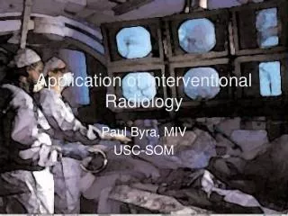
Application of Interventional Radiology Paul Byra, MIV USC-SOM
Angioplasty w/ vascular stenting Angiography Embolization CTA Cryotherapy Endovenous ablation of varicosities MRA MR guided biopsies Radiofrequency ablations of tumors Thrombolysis Transjugular Intrahepatic Portosystemic Shunt (TIPS) Ultrasound guided biopsies Uterine fibroid embolization Vascular access procedures Vertebroplasty Biliary drainage and stenting Fallopian tube catherization Gastrostomy tube insertion Urinary tract obstruction Procedures Performed And many more…
Case 1 • 60 y.o male w/ hx sig. for HTN, NIDDM, and 80 pack year smoking hx who presents with signs of claudication • Bilateral femoral, right popliteal, dorsalis pedis, and posterior tibial pulses present • No palpable popliteal, dorsalis pedis, posterior tibial pulses present on the left
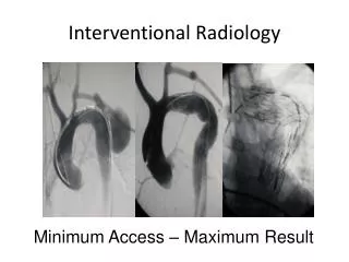
Catheter Angiography • Image blood vessels for disease, narrowing, occlusion, or aneurysms • Catheter is threaded through the vascular system to area of interest • Contrast is injected and visualized radiographically
Catheter Angiography Left common iliac stenosis Not pictured superficial femoral and proximal popliteal artery occlusion
Catheter Angiography Left iliac angioplasty and stent placement
Catheter Angiography Post stent placement
Case 2 • 30 y/o female with 16 week sized uterus • Negative UPT • Ultrasound significant for uterine fibroids • MRI of uterus • White arrows indicate the fibroids
Uterine Fibroid Emoblization • Arteriogram is performed, identifying fibroid vessels • Embolization is performed on one side until near stasis is achieved, then the other side is embolized • Normal myometrial vessles are spared
Uterine Fibroid Emoblization • 3 months S/P embolization • Uterine volume decreased by 66%
Case 3 • 50 y/o male with hx of Chronic Hepatitis and portal hypertension causing severe variceal bleeding • Patient is on beta-blocker and has failed sclerotherapy
TIPS • A catheter is placed in the right jugular vein • The catheter is threaded through the superior and inferior vena cava to the hepatic vein • Wall of the hepatic vein is punctured and the needle is directed across an approximate 2 inch gap to the portal vein. • Successful passage into the portal vein is determined by the pattern of dye injected through the catheter
TIPS • A guide wire is threaded through the needle to maintain the passage between the hepatic and portal veins.
TIPS • A balloon may be used across the passage to widen the holes in the vessel walls and the passage through the liver tissue
TIPS • Two stents are then positioned along the passage, overlapping in the liver tissue and extending into both veins. • The stents are opened to their maximum width with balloon dilation • Blood flow from the portal vein across the stents to the hepatic vein and on to the vena cava is confirmed with dye injection.
Case 4 • 72 y.o female with history significant for HTN, hyperlipidemia, and DM develops in the early afternoon a sudden, severe headache that is followed by left sided weakness of the arm and leg • Patient arrives to the ER 3 hours post-onset of symptoms • CT indicates nonhemorrhagic stroke • Patient’s BP 150/90, plt count >100k, normal bld glc; w/o history of intracranial hem., recent stroke or head trauma, recent MI, anticoagulation therapy, major surgery w/in past 14 days, or seizure during stroke • Basically no contraindications to tPA therapy but…it has been over 3 hours…hmmm…
Thrombolysis • When therapy cannot be initiated within three hours or when treatment with tPA during the first three hours is not sufficient to dissolve the blood clot, interventional neuroradiologists can provide intra-arterial thrombolysis treatment • Using x-ray guidance, an IR will insert a catheter through the femoral artery to the artery where the clot is present and will place the thrombolytic drug directly on the clot or break up the clot mechanically • When given locally this way, the tPA can be administered up to six hours after the onset of stroke symptoms
Thrombolysis • CTA shows cutoff of right middle cerebral artery CT perfusion images show decreased cerebral blood volume and cerebral blood flow with mean transit time representing ischemia.
Thrombolysis • Initial angiogram shows the middle cerebral artery cutoff • Road map angiogram shows the microcatheter crossing the clot before administration of tPA and microwire manipulation
Thrombolysis • At admission • Post thrombolysis angiogram
References • Society of Interventional Radiology. SIRweb.org • “Transjugular Intrahepatic Portosystemic Shunt”. University of Michigan Gastroenterology. http://www.med.umich.edu/1libr/aha/umliver09.htm • Interventional Neuroradiology. Massachusetts General Hospital. http://www.mgh-interventional-neurorad.org • Peripheral Vascular Disease. University of Toronto. http://surgclerk.med.utoronto.ca/