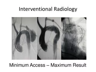
VASCULAR INTERVENTIONAL RADIOLOGY
VASCULAR INTERVENTIONAL RADIOLOGY. VASCULAR INTERVENTIONAL RADIOLOGY. Used as therapy of choice for the ff: Embolization of arteriovenous fistulas (AVFs) Ablation of renal function Treatment of testicular vein and ovarian vein varices Treatment of high-flow priaprism.

VASCULAR INTERVENTIONAL RADIOLOGY
E N D
Presentation Transcript
VASCULAR INTERVENTIONAL RADIOLOGY • Used as therapy of choice for the ff: • Embolization of arteriovenous fistulas (AVFs) • Ablation of renal function • Treatment of testicular vein and ovarian vein varices • Treatment of high-flow priaprism
TRANSCATHETER EMBOLIZATION • Embolization is the interventional radiological procedure (performed by an interventional radiologist) in which abnormal vessels are closed off with various substances (e.g., alcohol, glue, coil). • Embolization is a way of occluding (closing) one or more blood vessels that are doing more harm than good. Various materials may be used, depending on whether vessel occlusion is to be temporary or permanent, or whether large or small vessels are being treated.
TRANSCATHETER EMBOLIZATION • Renal Arteriovenous Fistulas and Malformations • Bleeding Sites • Tumors • Ablation of Renal Function • Embolization of Primary Varicocele • Treatment of High Flow Priapism
Renal Arteriovenous Fistulas and Malformations • Transcatheterembolization is the treatment of choice • May be congenital, spontaneous, acquired • Iatrogenic AVFs • Type most commonly treated • Occur as complications of other procedures (percutaneous renal biopsy, nephrostomy placement, pyelolithotomy)
Renal Arteriovenous Fistulas and Malformations • Classical angiographic finding of spontaneous or acquired AVF: • feeding artery with an early draining vein • Ancillary findings: • pseudoaneurysm and extravasation of contrast material • Congenital AVFs (AV malformation): • group of multiple coiled communicating vessels that may be associated with enlarged feeding arteries and draining veins
Renal Arteriovenous Fistulas and Malformations • Clinical Manifestation • Hematuria • Retroperitoneal or intraperitoneal hemorrhage • Congestive heart failure, cardiomegaly or both • Hypertension • Bruit • Duplex Doppler- most useful diagnostic procedure before angiographic intervention
Renal Arteriovenous Fistulas and Malformations Pre-embolization Post-embolization
Bleeding Sites • Transcatheter embolization plays a role in the management of hemorrhage originating in the kidney, ureter,, bladder and pelvis • Uses Gelfoams or coils
Gelfoam Slurry Preparation: First step is to cut the gelfoam into small pieces. Small pieces are then placed into a syringe and mixed with small amount of contrast solution utilizing a three-way stopcock. The obtained embolization material should be injected very carefully under fluoroscopic guidance using appropriate embolization materials (catheters, syringes etc.)
Metal coils. Occlusion occurs as a result of coil-induced thrombosis rather than mechanical occlusion of the lumen by the coil. To increase the thrombogenic effect, Dacron wool tails are attached to coils. The coils are available in many sizes and may be delivered through commonly used angiographic catheters
Tumors • Renal Cell Carcinoma • Transcatheterembolization is used as an adjunct to surgery • To reduce intraoperative hemorrhage • Allows immediate ligation of the renal vein • Palliation of nonresectable disease thatcauses pain and hematuria can be achieved by transcatheterembolization
Tumors • Gelfoam pledgets are used for preoperative embolization • Postembolization Syndrome (PES)- occurs hours after the procedure and may persist for days and is due to tissue necrosis caused by embolization • Severe pain - Fever • Nausea - Leukocytosis • Vomiting • Severity of PES is related to the quantity of infarcted tissue
Tumors • Angiomyolipoma • Selective embolization- effective method of controlling hemorrhage from benign renal lesions while preserving normal parenchyma • CT scan- identify the fat component of the tumor; used before embolization and for follow-up
Tumors • Ethanol- mixed with ionized oil then injected into the catheter to produce permanent occlusion of the vascular bed • Ionized oil- radiopaque and is useful for visualizing the flow of the embolic material during embolization
Ablation of Renal Function • Total renal infarction using transcatheterembolization is indicated in the ff: • Abolition of urine production to assist in healing or palliation of patients with urinary fistulas • Prevention of excessive proteinuria • Management of uncontrollable hypertension • Obstructive uropathy in patients who are poor surgical candidates
Ablation of Renal Function • Total renal ablation must be achieved so that perfusion of surviving parenchyma via pericapsular branches cannot occur • Ethanol- embolic agent of choice • PES may also occur managed with antibiotics and analgesics • Complication: embolization of adrenal artery • Avoided by using the occlusion balloon technique
Embolization of Primary Varicocele • Most varicoceles are left-sided • Color Doppler sonography is done before embolization • Equally effective comparing it to surgery • Performed with conscious intravenous sedation and local anesthesia • Transjugular venous approach- preferred by physicians
Embolization of Primary Varicocele • Embolization is achieved by placing coils within the gonadal vein commencing at the region of the inguinal ligament and continuing toward the renal vein until the gonadal vein and collateral vessels are occluded • 4% recurrence of varicocele after embolization • Minor complications: contrast extravasation, nontargetembolization, venospasm, hematoma
Treatment of High-Flow Priapism • A condition resulting from increased arterial flow into the lacunar spaces of the cavernous tissue • Color Doppler sonography demonstrates the abnormality • Nonselective transfemoral pelvic angiogram is performed to demonstrate the fistula, which originates from the pudendal artery • Followed by superselective catheterization of the injured artery using microcatheter • Fistula is closed by deploying microcoils
RENAL ARTERY ANGIOPLASTY AND STENTING • Ischemic nephropathy due to atherosclerotic vascular disease and renal artery stenosis - leading cause of progressive renal failure • Renal artery stenosis – most common cause of secondary hypertension • Ostial stenosis: within 3mm of the aortic lumen and is typical of atherosclerotic vascular disease • Non-ostial stenosis: seen in fibromuscular dysplsia • Branch vessel stenosis: seen in fibromuscular dysplsia