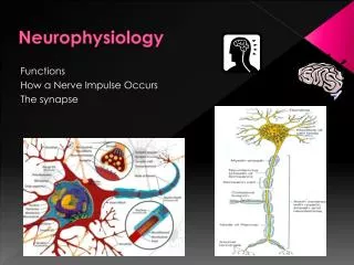
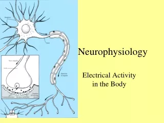
Neurophysiology
Neurophysiology . The Central Nervous System. Nervous System. Functions Sensory input – monitoring stimuli occurring inside & outside the body Integration – interpretation of sensory input Motor output – response to stimuli by activating effector organs. Figure 11.1.

Neurophysiology
E N D
Presentation Transcript
Neurophysiology The Central Nervous System
Nervous System • Functions • Sensory input – monitoring stimuli occurring inside & outside the body • Integration – interpretation of sensory input • Motor output – response to stimuli by activating effector organs Figure 11.1
Organization of the Nervous System • Central nervous system (CNS) • Brain and spinal cord • Integration and command center • Peripheral nervous system (PNS) • Paired spinal and cranial nerves • Carries messages to and from the spinal cord and brain
Peripheral Nervous System: Afferent Division • Afferent (sensory) division – transmits impulses from receptors to the CNS. • Somatic afferent fibers – carry impulses from skin, skeletal muscles, and joints • Visceral afferent fibers – transmit impulses from visceral organs
Peripheral Nervous System: Efferent Division • Motor (efferent) division – transmits impulses from the CNS to effector organs. Two subdivisions: • Somatic nervous system – provides conscious control of skeletal muscles • Autonomic nervous system – regulates smooth muscle, cardiac muscle, and glands
Sensory • General somatic senses – receptors are widely spread • Touch • Pain • Vibration • Pressure • Temperature • Proprioceptive senses – detect stretch in tendons and muscle • Body sense – position and movement of body in space • Special somatic senses • Hearing • Balance • Vision • Smell • Visceral sensory • General visceral senses – stretch, pain, temperature, nausea, and hunger • Widely felt in digestive and urinary tracts, and reproductive organs • Special visceral senses - taste
Motor • General somatic motor – signals contraction of skeletal muscles • Under our voluntary control • Often called “voluntary nervous system” • Visceral motor • Regulates the contraction of smooth and cardiac muscle • Makes up autonomic nervous system • Controls function of visceral organs • Often called “involuntary nervous system” • Autonomic nervous system
Central Nervous System: • Brain • Spinal cord
Organization of the Nervous System Figure 8-1: Organization of the nervous system
Nerve Tissue • The two principal cell types of the nervous system are: • Neurons – excitable cells that transmit electrical signals • Neuroglia - supporting cells
Neuron Classification • Functional: • Sensory (afferent) — transmit impulses toward the CNS • Motor (efferent) — carry impulses away from the CNS • Interneurons (association neurons) — shuttle signals through CNS pathways
Neuroglia Figure 12.6
CNS Protection • Hair, skin, cranium • Meninges • Cerebrospinal fluid • Blood brain barrier
Meningeal Layers • Meningeal layer of the brain cushion and protect delicate neural tissue Figure 9-4b
Cerebrospinal Fluid • Shock absorbing medium • Provides a optimum and stable environment for generating nerve impulses • Provides a medium for the exchange of nutrients and wastes between blood and nervous tissue
Cerebrospinal Fluid • Formed by selective transport across ependymal cells • Volume 125-150 ml and is replaced > 3 times/day, flow maintained by 10 mmHg pressure gradient • Path: ventricles subarachnoid space, reabsorbed into blood in dural sinuses through arachnoid villi
Blood Brain Barrier • Extensive capillaries & sinuses • Tight junctions promoted by astrocyte • Limits permeability for most molecules exceptO2, CO2, alcohol, steroids, H2O • Protects brain: hormones & circulating chemicals • Protects CNS from chemical fluctuations • Prevents entry of harmful substances • Prevents entry of molecules that could act as neurotransmitters • Brain receives 15% of blood pumped by heart • Brain responsible for about half of body’s glucose consumption • Membrane transporters move glucose from plasma into the brain interstitial fluid Figure 9-6: The blood-brain barrier
Brain Organization • Trillion interneurons fill the brain • Up to 200,000 synapses each • Levels of complexity • Cerebral cortex • Basal nuclei • Thalamus • Hypothalamus • Cerebellum • Brain stem
Brain component Cerebral cortex Cerebral cortex Basal nuclei (lateral to thalamus) Basal nuclei Thalamus (medial) Thalamus Hypothalamus Hypothalamus Cerebellum Cerebellum Midbrain Brain stem (midbrain, pons, and medulla) Brain stem Pons Medulla Spinal cord
Major Functions 1. Sensory perception 2. Voluntary control of movement 3. Language 4. Personality traits 5. Sophisticated mental events, such as thinking memory, decision making, creativity, and self-consciousness 1. Inhibition of muscle tone 2. Coordination of slow, sustained movements 3. Suppression of useless patterns of movements 1. Relay station for all synaptic input 2. Crude awareness of sensation 3. Some degree of consciousness 4. Role in motor control 1. Regulation of many homeostatic functions, such as temperature control, thirst, urine output, and food intake 2. Important link between nervous and endocrine systems 3. Extensive involvement with emotion and basic behavioral patterns 1. Maintenance of balance 2. Enhancement of muscle tone 3. Coordination and planning of skilled voluntary muscle activity 1. Origin of majority of peripheral cranial nerves 2. Cardiovascular, respiratory, and digestive control centers 3. Regulation of muscle reflexes involved with equilibrium and posture 4. Reception and integration of all synaptic input from spinal cord; arousal and activation of cerebral cortex 5. Role in sleep-wake cycle Brain component Cerebral cortex Basal nuclei Thalamus Hypothalamus Cerebellum Brain stem (midbrain, pons, and medulla)
Cerebrum • Highly developed • Makes up about 80% of total brain weight (largest portion of brain) • Inner core houses basal nuclei • Outer surface is highly convoluted cerebral cortex • Highest, most complex integrating area of the brain • Plays key role in most sophisticated neural functions
Cerebral Cortex • Three specializations • Sensory areas - sensory input translated into perception • Motor areas - direct skeletal muscle movement • Association areas - integrate information from sensory and motor areas, can direct voluntary behaviors
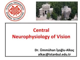
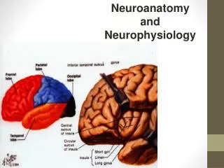
Cerebral Cortex • Each half of cortex divided into four major lobes • Occipital lobe - carries out initial processing of visual input • Temporal lobe - initial reception of sound sensation, taste, smell • Parietal lobe - somatosensory processing • Frontal lobe responsible for • Voluntary motor activity • Speaking ability • Elaboration of thought
Primary Somatosensory Cortex • Located in the postcentral gyrus, this area: • Receives information from the skin and skeletal muscles • Exhibits spatial discrimination • Somatosensory homunculus – caricature of relative amounts of cortical tissue devoted to each sensory function
Primary Motor Cortex • Located in the precentral gyrus • Composed of pyramidal cells whose axons make up the corticospinal tracts • Allows conscious control of precise, skilled, voluntary movements • Motor homunculus – caricature of relative amounts of cortical tissue devoted to each motor function
Language • Primary areas of cortical specialization for language • Broca’s area governs speaking ability • Wernicke’s area • Concerned with language comprehension • Responsible for formulating coherent patterns of speech Figure 9-23: Cerebral processing of spoken and visual language
Functional Areas of the Cerebral Cortex Figure 9-15
Brain Function: Cerebral Lateralization • Each lobe has special functions Figure 9-16
Cerebral Cortex • Schematic Linking of Various Regions of the Cortex
Basal Nuclei • Act by modifying ongoing activity in motor pathways • Primary functions • Regulates muscle tone throughout the body • Selecting and maintaining purposeful motor activity while suppressing useless or unwanted patterns of movement • Helping monitor and coordinate slow, sustained contractions, especially those related to posture and support • Controls large automatic movement
Thalamus • Final relay point for ascending sensory information • Coordinates the activities of the cerebral cortex and basal nuclei • Domain-specific information processing http://www.driesen.com/diencephalon.htm
Hypothalamus • Receives indirect sensory inputs from all sensory systems • Sends neural outputs to various motor control nuclei • Sends neural outputs to sympathetic and parasympathetic nervous systems • Sends both neural and hormonal outputs to pituitary
Hypothalamus • Controls somatic motor activities at the subconscious level • Controls autonomic function • Coordinates activities of the endocrine and nervous systems • Secretes hormones • Produces emotions and behavioral drives • Coordinates voluntary and autonomic functions • Regulates body temperature • Coordinates circadian cycles of activity • 4Fs: feeding, fighting, fleeing, and reproductive behavior
Limbic System • Cingulated gyrus • Coordinates sensory input with emotions • Emotional responses to pain • Basic, inborn behavioral patterns related to survival and perpetuation of the species • Regulates aggressive behavior • Hippocampus - sends memories out to the appropriate part of the cerebral hemisphere for long-term storage and retrieving them when necessary, Plays important role in motivation and learning • Amygdala - involved in emotional responses, hormonal secretions, and memory,
Cerebellum • Basic functions: coordination, balance, motor learning, etc. • Vestibulocerebellum – balance and control of eye movement • Spinocerebellum – enhances muscle tone and coordinates skilled voluntary movement – important in synchronization and timing • Receives input concerning desired action from motor cortex • Receives feedback concerning actual action from proprioceptors, vestibular apparatus, eyes • Compares inputs and sends adjustments or corrective signals to motor tracts • Cerebrocerebellum – planning and initiation of voluntary activity by providing input to the cortical motor areas also involved in procedural memories
Brain Stem: Midbrain, Pons & Medulla • An important link between spinal cord and higher brain levels, relays motor and sensory impulses between other “higher” parts of the brain and spinal cord • Midbrain – eye movement control • Pons/Medulla • Signal relay • Involuntary functions • Many cranial nerves enter • Pyramids – nerve tracts crossover
Cranial Nerves Table 9-1: The Cranial Nerves
Pons • Sensory and motor nuclei for four cranial nerves • Nuclei that help control respiration • Nuclei and tracts linking the cerebellum with the brain stem, cerebrum and spinal cord
Medulla oblongata • Contains relay stations and reflex centers • Cardiovascular and respiratory rhythmicity centers • Cardiovascular center - regulates rate and force of heartbeat and vasoconstriction/dilation • Respiratory center - regulates basic breathing rhythm • Reticular formation begins in the medulla oblongata and extends into more superior portions of the brainstem
Reticular Activating System • Network in brain stem • Arousal, sleep, pain, & muscle tone • Ascending fiber sends signals upward • Arouses and activates cerebral cortex • Controls overall degree of cortical alertness or level of consciousness: • maximum alertness • wakefulness • sleep • coma
ElectroEncephaloGram -EEG • Records electrical activity within cerebral cortex from EPSP, IPSPs • Used for • Diagnose cerebral dysfunction • Brain death • Sleep Patterns
Functions of Sleep • “Catch up” time – restore biochemical and physiological processes • Role of adenosine • Increased levels while awake • Inactivates RAS • Caffeine blocks adenosine receptors • Shift gears – long term structural and chemical changes required to consolidate memory and learning
Learning and Memory • Learning has two broad types • Associative – conditioning, linking two events together • Nonassociative • Habituation • Sensitization • Memory has several types • Short-term and long-term • Reflexive and declarative
Memory • Storage of acquired knowledge for later recall • Memory trace - Neural change responsible for retention or storage of knowledge • Short-term memory - Lasts for seconds to hours • Long-term memory - Retained for days to years • Consolidation - Process of transferring and fixing short-term memory traces into long-term memory stores • Working memory - Temporarily holds and interrelates various pieces of information relevant to a current mental task
Short Term Memory • Seconds to hours • Limited capacity • Rapid retrieval • Synaptic alterations • Changes in ion channels • Presynaptic facilitation cAMP • Long term potentiation (LTP)
Basic Learning - Behavior • Habituation – decreased responsiveness to stimulus • Closure of Ca++ channels leads • Reduced neurotransmitter release • Decrease EPSP • Sensitization – increase responsiveness • Release of serotonin from interneuron • Increases cAMP in presynaptic neuron • Blocks K+ channels and prolongs AP • Ca++ channels are open longer • Increasing neurotransmitter output
LTP • Glutamate acts as NT, binds to: • AMPA receptors elicit EPSPs • NMDA receptors open Ca++ channels > 2nd messenger system • Increases # of AMPA receptors / releases nitric oxide (NO) • NO causes presynaptic neuron to increase neurotransmitter release