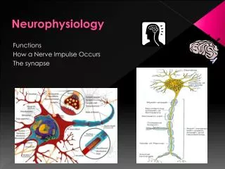
General Neurophysiology
General Neurophysiology. Axonal transport Transduction of signals at the cellular level Classification of nerve fibres. Olga Vajnerová, Department of physiology, 2nd Medical School Charles University Prague. Axonal transport. (axoplasmatic transport) Anterograde

General Neurophysiology
E N D
Presentation Transcript
General Neurophysiology Axonal transport Transduction of signals at the cellular level Classification of nerve fibres Olga Vajnerová, Department of physiology, 2nd Medical School Charles University Prague
Axonal transport (axoplasmatic transport) Anterograde Proteosynthesis in the cell body only (ER, Golgi apparatus) Retrograde Moving the chemical signals from periphery
Anterogradeaxonal transportfast (100 - 400 mm/day)MAP kinesin/mikrotubulesmoves neurotransmittersin vesicles and mitochondriaslow (0,5 – 10 mm/day)unknown mechanism structural components (cytoskeleton - aktin, myosin, tubulin), metabolic componentsRetrograde axonal transportfast (50 - 250 mm/day) MAP dynein/ mikrotubulesold mitochondria, vesicles (pinocytosis, receptor-mediated endocytosis in axon terminals, transport of e.g. growths factors),
Axonal transport in the pathogenesis of diseases Rabies virus (madness, hydrofobia) Replicates in muscle cell Axon terminal (endocytosis) Retrograde transport to the cell body Neurons produce copies of the virus CNS – behavioral changes Neurons innervating the salivary glands (anterograde transport) Tetanus toxin (produced by Clostridium tetani) Toxin is transported retrogradely in nerve cells Tetanus toxin is released from the nerve cell body Taken up by the terminals of neighboring neurons http://cs.wikipedia.org/wiki/Vzteklina
Axonal transport as a research tool Tracer studies (investigation of neuronal connections) Anterogradeaxonal transport Radioactively labeled amino acids (incorporated into proteins, transported in an anterograde direction, detectedby autoradiography) Injection into a group of neuronal cell bodies can identify axonal distribution Retrograde axonal transport Horseradish peroxidase is injected into regions containing axon terminals. Is taken up and transported retrogradely to the cell body. After histology preparationcan be visualized. Injection to axon terminalscan identify cell body
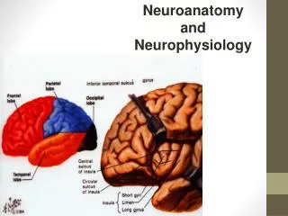
Transduction of signals at the cellular level Somatodendritic part – passive conduction of the signal, with decrement Axonal part –action potential, spreading without decrement, all-or-nothing law
Resting membrane potential Every living cell in the organism
Membrane potentialis not a potential. It is a difference of two potentials so itis a voltage, in fact.
K+ - - - + Ai + + + + When the membrane would be permeable for K+ only • K+ escapes out of the cell along concetration gradient • A- cannot leave the cell • Greater number of positive charges is on the outer side of the membrane K+ Na+ Cl-
Transduction of signals at the cellular level Axonal part –action potential, spreading without decrement, all-or-nothing law
Axon – the signal is carried without decrement Threshold All or nothing law
Action potential Membrane conductance for Na+ a pro K+
Transduction of signals at the cellular level Somatodendritic part – passive conduction of the signal, with decrement
Dendrite and cell body – signal is propagated with decrement
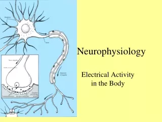
Origin of the electrical signal electrical stimulus sensory input neurotransmitter on synapses
Axonal part of the neuronAP – voltage-gated Ca2+ channels –neurotransmitter release Arrival of an AP in the terminal opens voltage-gated Ca2+ channels, causing Ca2+ influx, which in turn triggers transmitter release.
Somatodendritic part of neuron Receptors on the postsynaptic membrane • Excitatory receptors open Na+, Ca2+channelsmembrane depolarization • Inhibitoryreceptors open K+, Cl-channels membrane hyperpolarization • EPSP – excitatory postsynaptic potential • IPSP – inhibitory postsynaptic potential
Summation of signals spatial and temporal
Potential changes in the area of trigger zone (axon hillock) • Interaction of all synapses • Spatial summation – currentsfrom multiple inputs add algebraically up • Temporal summation –if another APsarrive at intervals shorter than the duration of the EPSP Trigger zone
Transduction of signals at the cellular level EPSP IPSP Initial segment AP Ca2+ influx Neurotransmitter Neurotransmitter releasing
EPSP IPSP Neuronal activity in transmission of signals Discharge configurationsof various cells
1.AP, activation of the voltage-dependent Na+ channels (soma, area of the initial segment) 2. ADP, after-depolarization, acctivation of a high threshold Ca2+ channels, localized in the dendrites 3.AHP, after-hyperpolarization, Ca2+ sensitive K+ channels 4.Rebound depolarization, low threshold Ca2+ channels, (probably localized at the level of the soma Influence of one cell on the signal transmission Threshold RMP Hammond, C.:Cellular and Molecular Neurobiology. Academic Press, San Diego 2001: str. 407.
Origin of the electrical signalelectrical stimulussensory inputneurotransmitter on synapses
Sensory input Sensory transduction – conversion of stimulus from the external or internal environment into an electrical signal Phototransduction Chemotransduction Mechanotransduction Signals: sound wave (auditory), taste, light photon (vision), touch, pain, olfaction, muscle spindle,
Sensory input Sensory transduction – conversion of stimulus from the external or internal environment into an electrical signal Phototransduction light photon (vision), Chemotransduction taste,pain olfaction Mechanotransduction sound wave (auditory),touch,muscle spindle Osmoreceptors, thermoreceptors
The compound action potential Program neurolab Diferences between the velocities of individual fibres give rise to a dispersed compoud action potential
Two different systems are in use for classifying nerve fibres
Degeneration and regeneration in the nervous system Myelin sheath of axons in PNS(a membranous wrapping around the axon)
Myelin sheath of axons in PNS(a basal lamina) Basal lamina
Injury of the axon in PNS • Compression, crushing, cutting – degeneration of the distal axon - but the cell body remains intact (Wallerian degeneration, axon is removedby macrophages) • Schwann cells remainand their basal lamina (band of Büngner) • Proximal axon sprouts (axonal sprouting) • Prognosis quo ad functionem • Compression, crushing –good, Schwann cells remain in their original orientation, axons can find their original targets • Cutting – worse, regeneration is less likely to occure
Injury of the axon in CNS • Oligodendrocytes do not create a basal lamina and a band of Büngner • Regeneration to a functional state is impossible Trauma of the CNS • proliferation and hypertrophy of astrocytes, astrocytic scar
Injury of the axon in PNS after amputation • Amputation of the limb • Proximal stumpfail to enter the Schwann cell tube, instead ending blindly in connective tissue • Blind ends rolle themselves into a ball and form aneuroma – phantom pain