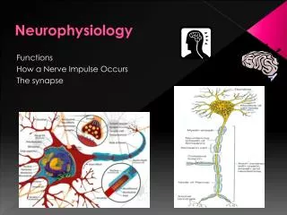
Neurophysiology
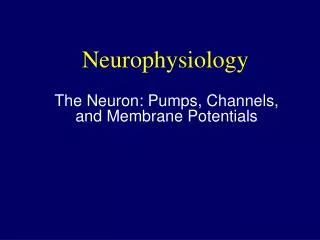
Neurophysiology. Physiological Psychology PSYC-465. What level(s) of analysis?. Behavior and Cognition. Systems and Circuits. Synapses and Neurons. Genes and Molecules. Membrane potential – the difference in electrical charge between the inside and outside of a cell.


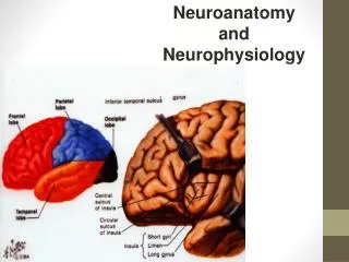
Neurophysiology
E N D
Presentation Transcript
Neurophysiology Physiological Psychology PSYC-465
What level(s) of analysis? Behavior and Cognition Systems and Circuits Synapses and Neurons Genes and Molecules
Membrane potential – the difference in electrical charge between the inside and outside of a cell. • The tip of one electrode is positioned outside the neuron. • The tip of a fine microelectrode (1/1,000 mm) is advanced until it pierces the membrane and is positioned inside the neuron. Recording the Membrane Potential Experimental setup to record a neuron’s membrane potential
Resting Membrane Potential • Resting potential is -70 mV • The inside of the neuron is 70 mV less than the outside (extracellular fluid). • The neuron is said to be polarized – a -70 mV charge is built up across the membrane.
Cl- Cl- Cl- Cl- Cl- Cl- Cl- Cl- Cl- Cl- A-- A-- A-- Na+ A-- A-- Na+ Na+ A-- Na+ Na+ Na+ K+ A-- K+ Na+ Na+ K+ K+ K+ K+ K+ Na+ K+ K+ • Ions are charged particles. There are more negative ions relative to positive ones inside the neuron. This is due to the interaction of 4 factors (2 act to distribute ions evenly and 2 are features of the cell membrane): • Random motion- particles in constant motion move down their concentration gradients. • Electrostatic pressure– like charges repel, opposites attract. • No single class of ions is distributed evenly across both sides of the cell membrane. • Sodium (Na+) and chloride (Cl-) are greater outside. • Potassium (K+) and large protien ions (Anions; A--) are greater inside. Ionic Basis of the Resting Potential
Cl- Cl- Cl- Cl- Cl- Cl- Cl- Cl- Cl- Cl- A-- A-- A-- Na+ A-- A-- Na+ Na+ A-- Na+ Na+ Na+ K+ A-- K+ Na+ Na+ K+ K+ K+ K+ K+ Na+ K+ K+ • 3)Differential permeability– The membrane has specialized pores called ion channels for each kind of ion. • Potassium (K+) and chloride (Cl-) ions pass readily through their channels. • Sodium (Na+) ions pass through with difficulty. • Large protien ions (A--) are trapped inside. • IF some ions can pass through the membrane then what prevents them from flowing down their concentration gradients? • IS it electrostatic pressure? Ionic Basis of the Resting Potential
Cl- 70 mV from CG Cl- Cl- Cl- Cl- Cl- Cl- Cl- 50 mV from CG Cl- Cl- A-- 70 mV from ES A-- A-- Na+ A-- A-- Na+ Na+ A-- Na+ Na+ Na+ K+ A-- K+ Na+ Na+ K+ K+ 70 mV from ES K+ K+ K+ Na+ K+ K+ 90 mV from CG 70 mV from ES To answer the question Hodgkin & Huxley calculated the amount of electrostatic charge that would be required for each ion that can pass through the membrane (Cl-, K+ and Na+) to move down their concentration gradients. Hodgkin & Huxley (1950)
Hodgkin & Huxley (1950) • Concluded that there are active mechanisms in the cell membrane to counteract the passive influx (inflow) of Na+ ions and the passive efflux (outflow) of K+ ions.
Na+ K+ Na+ K+ Na+ 4) Subsequently it was discovered that the sodium-potassium pump exchanges 3 sodium ions out of the neuron for every 2 potassium ions brought into the neuron. This is an active mechanism that requires energy (ATP) to maintain the resting membrane potential. Sodium-Potassium Pump
How do neurons talk to each other? • When one neuron fires an action potential, it causes the release of neurotransmitter molecules into the small space that separates the terminal bouton from the receptive portion of the neuron (e.g., a dendritic spine). The space is called the synapse. • Neurotransmitter molecules bind to receptors on the next neuron, causing one of two events.
Two Postsynaptic Events • Depolarization – decreases the resting membrane potential (e.g., from -70 to -67 mV). • Hyperpolarization – increases the resting membrane potential (e.g., from -70 to -72 mV).
Postsynaptic Potentials (PSPs) • Depolarizations are called Excitatory postsynaptic potentials (EPSPs) – they increase the likelihood that the postsynaptic (receiving) neuron will itself generate an action potential. • Hyperpolarizations are called Inhibitory postsynaptic potentials (IPSPs) – they decreases the likelihood that the postsynaptic neuron will generate an action potential.
Postsynaptic Potentials (PSPs) • Both EPSPs and IPSPs are graded events – i.e., the amplitudes of both PSPs are proportional to the intensity of the signal (they come in different sizes). • Weak signals generate small PSPs and strong signals elicit large PSPs.
Postsynaptic Potentials (PSPs) • PSPs travel passively from the site of origin, similar to the way an electrical signal travels through a cable. • Accordingly, PSPs travel fast but are decremental (i.e., they decrease in amplitude the further they travel).
Integration of PSPs and Generation of Action Potentials (APs) • In order to generate an AP (making a neuron “fire”) the threshold of excitation must be reached at the beginning section of the axon, near the axon hillock. • Integration of IPSPs and EPSPs must result in a potential of about -65mV in order to generate an AP
Integration • Adding or combining a number of individual signals into one overall signal. • Temporal summation – integration of events happening at different times. • Spatial summation - integration of events happening at different places.
Spatial Summation Local PSPs produced simultaneously on different parts of the neuron sum to produce greater PSPs or cancel each other out.
Temporal Summation PSPs produced in rapid succession at the same synapse sum to form a greater signal.
Comparison of PSPs and APs In contrast to PSPs, the AP is a massive mommentary reversal of the membrane potential from -70 mV to +50 mV.
Ionic Basis of the AP Cl- Na+ Cl- Na+ Cl- Na+ Na+ Na+ Na+ Cl- Na+ Cl- Na+ Na+ Na+ + + + + + + Sodium channel Sodium channel Sodium channel Potassium channel - - - - - - K+ A-- K+ K+ K+ K+ A-- A-- A-- A-- K+ K+ K+
Refractory Periods • Absolute refractory – a brief period (1-2 ms) after the initiation of an AP during which it is not possible to elicit another AP. • Relative refractory – the period in which it is possible to fire an AP, but only if higher-than-normal levels of stimulation are applied.
Refractory Periods Refractory periods are responsible for two characteristics of neural conduction: • APs normally travel in one direction. • Rate of neural firing is related to the intensity of the stimulation.
Saltatory Conduction Transmission of APs in myelinated axons – saltare (to “skip” or “jump”). • AP conduction along myelinated segments of an axon is passive. • i.e., it travels fast but gets weaker the farther it goes. • The signal is still strong enough to generate a full AP at the next node.
How fast are APs? Conduction speed depends on: • Diameter of axon (faster in large axons). • Myelination (faster in myelinated axons).
Conduction in Myelinated Axons • Passive movement of AP within myelinated portions occurs instantly • Nodes of Ranvier (unmyelinated) • Where ion channels are found • Where full AP is seen • AP appears to jump from node to node • Saltatory conduction • http://www.brainviews.com/abFiles/AniSalt.htm
The Changing View of Dendritic Function Three recently discovered characteristics of dendrites: • Some can generate APs that travel in either direction. • Dendritic spines restrict chemical changes to the immediate area of the synapse (they compartmentalize the dendrite). • Spines change rapidly (within minutes to hours) in shape and number in response to neural stimulation.
Overview of Neural signals • EPSPs and IPSPs (PSPs) are graded events initiated by the action of neurotransmitters binding to receptors on the postsynaptic membrane. • PSPs summate spatially and temporally • If EPSP signals are greater, causing a change in membrane potential (depolarization) to -64 mV (threshold of exitation) at the beginning segment of the axon, then the postsynaptic neuron will fire an AP • Once initiatiated the AP travels the full length of the axon to the terminal buttons.