Neurophysiology
Neurophysiology. Chapters 10-12. Control and Integration. Nervous system composed of nervous tissue cells designed to conduct electrical impulses rapid communication to specific cells or groups of cells Endocrine system composed of various tissue types


Neurophysiology
E N D
Presentation Transcript
Neurophysiology Chapters 10-12
Control and Integration • Nervous system • composed of nervous tissue • cells designed to conduct electrical impulses • rapid communication to specific cells or groups of cells • Endocrine system • composed of various tissue types • cell communication solely through chemical messengers • slow speed of action, broadcast
Nervous System Organization:Radial Symmetric Animals • Neural Net • Cnidarians and ctenophorans • Echinoderms • no specific CNS Fig 10.4
Nervous System Organization:Bilateral Symmetric Animals • Nerve Cords • Longitudinally oriented tracts of neurons, with lateral commissures • Evolutionary Trends • Reduction of nerve cord numbers • Cephalization – anterior concentration of nerve tissue (brain) Figs 10.4-10.6
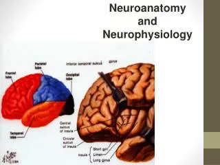
Nervous System Organization: Bilateral Symmetric Animals • Central Nervous System • Brain + Spinal Cord • control center (integration) • Peripheral Nervous System • cranial nerves and spinal nerves • connects CNS to sensory receptors, muscles and glands Fig 10.7
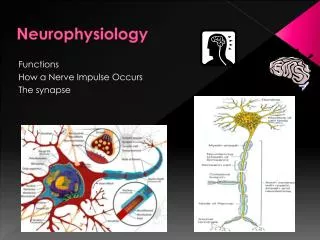
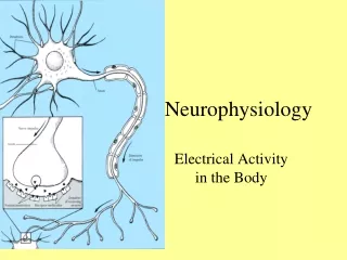
Cell Body nucleus and organelles Dendrites receive information Axon conduct electrical signals (action potentials) axon hillock - site where AP’s originate axon terminals - where chemical signals are released Neurons Fig 11.1
Membrane Potentials • All cell membranes are electrically polarized • Unequal distribution of charges • Membrane potential (mV) = difference in charge across the membrane • Due to unequal ion concentrations across cell membrane (fixed anions)
Ion Movements • K+ • [K+] higher inside cell than outside • Attracted to fixed anions inside cell • High membrane permeability • Flows slowly out of cell • Na+ • [Na+] higher outside cell than inside • Attracted to fixed anions inside cell • Low membrane permeability • Flows slowly into cell
Equilibrium Potential • Equilibrium (no net movement) will be reached when a particular electrical potential is reached • Equilibrium potential = theoretical electrical potential at which the net flow of ions across the membrane is 0 • balance between EG and CG is achieved
Equilibrium Potential • Equilibrium potential is calculated for a particular ion using the Nernst Equation Ex = RT/zF ln[Xo]/[Xi] • Ex = equilibrium potential (mV) • R = gas constant (8.31 J/(K*mol)) • T = temperature (K) • z = charge of the ion • F = Faraday’s constant (96500 coulombs/mole) • [Xo] and [Xi] = concentrations of ion “X” inside and outside the cell
Equilibrium Potential • For equilibrium potentials of Na+ and K+ in eutherian mammals (Tb = 310 K) Ex = 61 log [Xo]/[Xi] • Equilibrium potential for K+ (EK) = -90 mV • Equilibrium potential for Na+ (ENa) = +60 mV
Distribution of Inorganic Ions • Different ions unevenly distributed across cell membrane • Each has own specific equilibrium potential and membrane permeability Table 11.1
Resting Potentials • Resting potential • Typical membrane potential for cells • Depends on concentration gradients and membrane permeabilities for different ions involved • Goldman Equation • -65 to -85 mV (unequal to EKor ENa) • [Na+] and [K+] inside the cell are maintained using Na+/K+ pumps Na/Kpump ECF (+) ICF (-)
Electrical Activity of Neurons:Electrical Signals • Electrical signals • due to changes in membrane permeability and altering flow of charged particles • changes in permeability are due to changing the number of open membrane channels -70 mV -30 mV
Membrane Proteins Involved in Electrical Signals • Non-gated ion channels (leak channels) • always open • specific for a particular ion • Gated Ion channels • open only under particular conditions (stimulus) • voltage-gated, ligand-gated, stress-gated • Ion pumps • active (require ATP) • maintain ion gradients Figs 11.9, 11.12
Types of Electric Signals: Graded Potentials • occur in dendrites / cell body • small, localized change in membrane potential • change of only a few mV • opening of chemically-gated or physically-gated ion channels • travels only a short distance (few mm) + + + + + + + + + + + + -70 mV -55 mV -63 mV -70 mV -68 mV -70 mV - - - - - - - - - - See Fig 11.6
Types of Electric Signals: Graded Potentials • a triggered event • requires stimulus • e.g. light, touch, chemical messengers • graded • stimulus intensity → change in membrane potential + + + + + + + + + + + + - - - - - - - - - -
Graded Potential + + + + + + + + + + + + + + + + + + + + + + + + + + -60 mV -40 mV -50 mV -70 mV - - - - - - - - - - - - - - - - - - - -
0 mV -70 Types of Electric Signals:Action Potentials • begins at the axon hillock, travels down axon • brief, rapid reversal of membrane potential • Large change (~70-100 mV) • Opening of voltage-gated Na+ and K+ channels • self-propagating - strength of signal maintained • long distance transmission
Types of Electric Signals:Action Potentials • triggered • membrane depolarization (depolarizing graded potential) • "All or none" • axon hillock must be depolarized a minimum amount (threshold potential) • if depolarized to threshold, AP will occur at maximum strength • if threshold not reached, no AP will occur + + + + + + + + + + + + + + + + + + + + + + + + + + + + + + + + +
+30 0 mV threshold -70 Action Potential:Depolarization Action Potential: Repolarization • Na+ channels close • Delayed opening of voltage-gated K+ channels • K+ rushes out of the cell • membrane potential restored • voltage-gated Na+ channels open • Na+ enters cell → further depolarization → more channels open → further depolarization • membrane reverses polarity (+30 mV) • K+ channels close • [Na+] and [K+] restored by the Na+-K+ pump • Triggering event (graded potential) causes membrane to depolarize • slow increase until threshold is reached
Action Potential Propagation • Na+ moving into one segment of the neuron quickly moves laterally inside the cell • Depolarizes adjacent segment to threshold Fig 11.23
Conduction Velocity • Conduction velocity • speed at which the action potential travels down the length of an axon • dictates speed of response • Velocity directly related to axon diameter • Increased diameter lowers internal resistance to ion flow • V α √ D in unmyelinated axons • V α D in myelinated axons Fig 11.24
Action Potential Propagation:Myelinated Axons • myelin - lipid insulator • membranes of certain glial cells • Nodes of Ranvier contain lots of Na+ channels • Saltatory conduction • signals “jump” from one node to the next • AP conduction speed 50-100x • Vertebrates tend to have more myelinated axons than invertebrates Fig 11.25
Chemical Synapses • presynaptic neuron • synaptic terminal bouton • contains synaptic vesicles filled with neurotransmitter • synaptic cleft • space in-between cells • postsynaptic neuron • subsynaptic membrane • contains receptors that bind neurotransmitter Fig 11.1
Chemical Synapses + - - + • Many voltage-gated Ca2+ channels in the terminal bouton • AP in knob opens Ca2+ channels • Ca2+ rushes in. • Ca2+ induced exocytosis of synaptic vesicles • Transmitter diffuses across synaptic cleft and binds to receptors on subsynaptic membrane + - - + + - - + Ca2+ Ca2+ + + - - Ca2+ Ca2+ Ca2+ Ca2+ Ca2+ Ca2+ Calmodulin Protein Kinase C Synapsins
Chemical Synapses • Generate Postsynaptic Potentials • Specific ion channels in subsynaptic membrane open, altering membrane permeability • If depolarizing graded potential is strong enough to reach threshold - generates action potential in postsynaptic cell • Metabotropic actions • Long lasting effects (e.g., synaptic changes in learning and memory) Figs 12.7, 12.18, 12.20, 12.22
Types of Postsynaptic Potentials • excitatory postsynaptic potentials (EPSPs) • Transmitter binding opens Na+ channels in the postsynaptic membrane • Small depolarization of postsynaptic neuron • More positive inside the cell • closer to threshold Fig 12.5
Types of Postsynaptic Potentials • inhibitory postsynaptic potentials (IPSPs) • Transmitter binding opens K+ or Cl- ion channels • K+ flows out or Cl- flows in down gradients • Small hyperpolarization of postsynaptic neuron • More negative inside cell • further from threshold Fig 12.5
Summation • spatial summation • numerous presynaptic fibers may converge on a single postsynaptic neuron • additive effects of numerous neurons inducing EPSPs and IPSPs on the postsyn. neuron • temporal summation • additive effects of EPSPs and IPSPs occurring in rapid succession • next synaptic event occurs before membrane recovers from previous event Fig 12.5