Salivary Gland Neoplasms
790 likes | 1.83k Vues
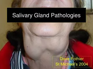
Salivary Gland Neoplasms. Benign NeoplasmsMalignant NeoplasmsControversial Issues. Salivary Gland Neoplasms. Diverse histopathologyRelatively uncommon2% of head and neck neoplasmsDistributionParotid: 80% overall; 80% benignSubmandibular: 15% overall; 50% benignSublingual/Minor: 5% overall; 40% benign.

Salivary Gland Neoplasms
E N D
Presentation Transcript
1. Salivary Gland Neoplasms Elizabeth J. Rosen, MD
Shawn D. Newlands, MD, PhD
6/26/02
2. Salivary Gland Neoplasms Benign Neoplasms
Malignant Neoplasms
Controversial Issues
3. Salivary Gland Neoplasms Diverse histopathology
Relatively uncommon
2% of head and neck neoplasms
Distribution
Parotid: 80% overall; 80% benign
Submandibular: 15% overall; 50% benign
Sublingual/Minor: 5% overall; 40% benign
4. Pleomorphic Adenoma Most common of all salivary gland neoplasms
70% of parotid tumors
50% of submandibular tumors
45% of minor salivary gland tumors
6% of sublingual tumors
4th-6th decades
F:M = 3-4:1
5. Pleomorphic Adenoma Slow-growing, painless mass
Parotid: 90% in superficial lobe, most in tail of gland
Minor salivary gland: lateral palate, submucosal mass
Solitary vs. synchronous/metachronous neoplasms
6. Pleomorphic Adenoma Gross pathology
Smooth
Well-demarcated
Solid
Cystic changes
Myxoid stroma
7. Pleomorphic Adenoma Histology
Mixture of epithelial, myopeithelial and stromal components
Epithelial cells: nests, sheets, ducts, trabeculae
Stroma: myxoid, chrondroid, fibroid, osteoid
No true capsule
Tumor pseudopods
8. Pleomorphic Adenoma
9. Pleomorphic Adenoma Treatment: complete surgical excision
Parotidectomy with facial nerve preservation
Submandibular gland excision
Wide local excision of minor salivary gland
Avoid enucleation and tumor spill
10. Warthin�s Tumor AKA: papillary cystadenoma lymphomatosum
6-10% of parotid neoplasms
Older, Caucasian, males
10% bilateral or multicentric
3% with associated neoplasms
Presentation: slow-growing, painless mass
11. Warthin�s Tumor Gross pathology
Encapsulated
Smooth/lobulated surface
Cystic spaces of variable size, with viscous fluid, shaggy epithelium
Solid areas with white nodules representing lymphoid follicles
12. Warthin�s Tumor Histology
Papillary projections into cystic spaces surrounded by lymphoid stroma
Epithelium: double cell layer
Luminal cells
Basal cells
Stroma: mature lymphoid follicles with germinal centers
13. Warthin�s Tumor
14. Oncocytoma Rare: 2.3% of benign salivary tumors
6th decade
M:F = 1:1
Parotid: 78%
Submandibular gland: 9%
Minor salivary glands: palate, buccal mucosa, tongue
15. Oncocytoma Presentation
Enlarging, painless mass
Technetium-99m pertechnetate scintigraphy
Mitochondrial hyperplasia
16. Oncocytoma Gross
Encapsulated
Homogeneous, smooth
Orange/rust color
Histology
Cords of uniform cells and thin fibrous stroma
Large polyhedral cells
Distinct cell membrane
Granular, eosinophilic cytoplasm
Central, round, vesicular nucleus
17. Oncocytoma Electron microscopy:
Mitochondrial hyperplasia
60% of cell volume
18. Monomorphic Adenomas Basal cell, canalicular, sebaceous, glycogen-rich, clear cell
Basal cell is most common: 1.8% of benign epithelial salivary gland neoplasms
6th decade
M:F = approximately 1:1
Caucasian > African American
Most common in parotid
19. Basal Cell Adenoma Solid
Most common
Solid nests of tumor cells
Uniform, hyperchromatic, round nuclei, indistinct cytoplasm
Peripheral nuclear palisading
Scant stroma
20. Basal Cell Adenoma Trabecular
Cells in elongated trabecular pattern
Vascular stroma
21. Basal Cell Adenoma Tubular
Multiple duct-like structures
Columnar cell lining
Vascular stroma
22. Basal Cell Adenoma Membranous
Thick eosinophilic hyaline membranes surrounding nests of tumor cells
�jigsaw-puzzle� appearance
23. Monomorphic Adenomas Canalicular adenoma
7th decade
F:M � 1.8:1
Most common in minor salivary glands of the upper lip (74%)
Painless submucosal mass
24. Canalicular Adenoma Histology
Well-circumscribed
Multiple foci
Tubular structures line by columnar or cuboidal cells
Vascular stroma
25. Myoepithelioma <1% of all salivary neoplasms
3rd-6th decades
F>M
Minor salivary glands > parotid > submandibular gland
Presentation: asymptomatic mass
26. Myoepithelioma Histology
Spindle cell
More common
Parotid
Uniform, central nuclei
Eosinophilic granular or fibrillar cytoplasm
Plasmacytoid cell
Polygonal
Eccentric oval nuclei
27. Mucoepidermoid Carcinoma Most common salivary gland malignancy
5-9% of salivary neoplasms
Parotid 45-70% of cases
Palate 18%
3rd-8th decades, peak in 5th decade
F>M
Caucasian > African American
28. Mucoepidermoid Carcinoma Presentation
Low-grade: slow growing, painless mass
High-grade: rapidly enlarging, +/- pain
**Minor salivary glands: may be mistaken for benign or inflammatory process
Hemangioma
Papilloma
Tori
29. Mucoepidermoid Carcinoma Gross pathology
Well-circumscribed to partially encapsulated to unencapsulated
Solid tumor with cystic spaces
30. Mucoepidermoid Carcinoma Histology�Low-grade
Mucus cell > epidermoid cells
Prominent cysts
Mature cellular elements
31. Mucoepidermoid Carcinoma Histology�Intermediate- grade
Mucus = epidermoid
Fewer and smaller cysts
Increasing pleomorphism and mitotic figures
32. Mucoepidermoid Carcinoma Histology�High-grade
Epidermoid > mucus
Solid tumor cell proliferation
Mistaken for SCCA
Mucin staining
33. Mucoepidermoid Carcinoma Treatment
Influenced by site, stage, grade
Stage I & II
Wide local excision
Stage III & IV
Radical excision
+/- neck dissection
+/- postoperative radiation therapy
34. Adenoid Cystic Carcinoma Overall 2nd most common malignancy
Most common in submandibular, sublingual and minor salivary glands
M = F
5th decade
Presentation
Asymptomatic enlarging mass
Pain, paresthesias, facial weakness/paralysis
35. Adenoid Cystic Carcinoma Gross pathology
Well-circumscribed
Solid, rarely with cystic spaces
infiltrative
36. Adenoid Cystic Carcinoma Histology�cribriform pattern
Most common
�swiss cheese� appearance
37. Adenoid Cystic Carcinoma Histology�tubular pattern
Layered cells forming duct-like structures
Basophilic mucinous substance Histology�solid pattern
Solid nests of cells without cystic or tubular spaces
38. Adenoid Cystic Carcinoma Treatment
Complete local excision
Tendency for perineural invasion: facial nerve sacrifice
Postoperative XRT
Prognosis
Local recurrence: 42%
Distant metastasis: lung
Indolent course: 5-year survival 75%, 20-year survival 13%
39. Acinic Cell Carcinoma 2nd most common parotid and pediatric malignancy
5th decade
F>M
Bilateral parotid disease in 3%
Presentation
Solitary, slow-growing, often painless mass
40. Acinic Cell Carcinoma Gross pathology
Well-demarcated
Most often homogeneous
41. Acinic Cell Carcinoma Histology
Solid and microcystic patterns
Most common
Solid sheets
Numerous small cysts
Polyhedral cells
Small, dark, eccentric nuclei
Basophilic granular cytoplasm
42. Acinic Cell Carcinoma Treatment
Complete local excision
+/- postoperative XRT
Prognosis
5-year survival: 82%
10-year survival: 68%
25-year survival: 50%
43. Adenocarcinoma Rare
5th to 8th decades
F > M
Parotid and minor
salivary glands
Presentation:
Enlarging mass
25% with pain or facial weakness
44. Adenocarcinoma Histology
Heterogeneity
Presence of glandular structures and absence of epidermoid component
Grade I
Grade II
Grade III
45. Adenocarcinoma Treatment
Complete local excision
Neck dissection
Postoperative XRT
Prognosis
Local recurrence: 51%
Regional metastasis: 27%
Distant metastasis: 26%
15-year cure rate:
Stage I = 67%
Stage II = 35%
Stage III = 8%
46. Malignant Mixed Tumors Carcinoma ex-pleomorphic adenoma
Carcinoma developing in the epithelial component of preexisting pleomorphic adenoma
Carcinosarcoma
True malignant mixed tumor�carcinomatous and sarcomatous components
Metastatic mixed tumor
Metastatic deposits of otherwise typical pleomorphic adenoma
47. Carcinoma Ex-Pleomorphic Adenoma 2-4% of all salivary gland neoplasms
4-6% of mixed tumors
6th-8th decades
Parotid > submandibular > palate
Risk of malignant degeneration
1.5% in first 5 years
9.5% after 15 years
Presentation
Longstanding painless mass that undergoes sudden enlargement
48. Carcinoma Ex-Pleomorphic Adenoma Gross pathology
Poorly circumscribed
Infiltrative
Hemorrhage and necrosis
49. Carcinoma Ex-Pleomorphic Adenoma Histology
Malignant cellular change adjacent to typical pleomorphic adenoma
Carcinomatous component
Adenocarcinoma
Undifferentiated
50. Carcinoma Ex-Pleomorphic Adenoma Treatment
Radical excision
Neck dissection (25% with lymph node involvement at presentation)
Postoperative XRT
Prognosis
Dependent upon stage and histology
51. Carcinosarcoma Rare: <.05% of salivary gland neoplasms
6th decade
M = F
Parotid
History of previously excised pleomorphic adenoma, recurrent pleomorphic adenoma or recurring pleomorphic treated with XRT
Presentation
52. Carcinosarcoma Gross pathology
Poorly circumscribed
Infiltrative
Cystic areas
Hemorrhage, necrosis
Calcification
53. Carcinosarcoma Histology
Biphasic appearance
Sarcomatous component
Dominant
chondrosarcoma
Carinomatous component
Moderately to poorly differentiated ductal carcinoma
Undifferentiated
54. Carcinosarcoma Treatment
Radical excision
Neck dissection
Postoperative XRT
Chemotherapy (distant metastasis to lung, liver, bone, brain)
Prognosis
Poor, average survival less than 2 � years
55. Squamous Cell Carcinoma 1.6% of salivary gland neoplasms
7th-8th decades
M:F = 2:1
MUST RULE OUT:
High-grade mucoepidermoid carcinoma
Metastatic SCCA to intraglandular nodes
Direct extension of SCCA
56. Squamous Cell Carcinoma Gross pathology
Unencapsulated
Ulcerated
fixed
57. Squamous Cell Carcinoma Histology
Infiltrating
Nests of tumor cells
Well differentiated
Keratinization
Moderately-well differentiated
Poorly differentiated
No keratinization
58. Squamous Cell Carcinoma Treatment
Radical excision
Neck dissection
Postoperative XRT
Prognosis
5-year survival: 24%
10-year survival: 18%
59. Polymorphous Low-Grade Adenocarcinoma 2nd most common malignancy in minor salivary glands
7th decade
F > M
Painless, submucosal mass
Morphologic diversity
Solid, glandular, cribriform, ductular, tubular, trabecular, cystic
60. Polymorphous Low-Grade Adenocarcinoma Histology
Isomorphic cells, indistinct borders, uniform nuclei
Peripheral �Indian-file� pattern
Treatment
Complete yet conservative excision
61. Clear Cell Carcinoma AKA glycogen-rich
Palate and parotid
6th-8th decade
M = F
Histology
Uniform, round or polygonal cells
Peripheral dark nuclei
Clear cytoplasm
Treatment
Complete local excision
62. Epithelial-Myoepithelial Carcinoma < 1% of salivary neoplasms
6th-7th decades, F > M, parotid
? Increased risk for 2nd primary
Histology
Tumor cell nests
Two cell types
Thickened basement membrane
Treatment
Surgical excision
63. Undifferentiated Carcinoma Lymphoepithelial
Eskimos: parotid, F > M, familial
Asian: submandibular, M > F
Large-cell
Bimodal peaks
M > F
Parotid
Small-cell
6th-7th decades
M:F = 1.6:1
parotid
64. Controversial Issues Management of the N0 Neck
Recurrence in the neck = low likelihood of salvage
Parotid: clinical neck disease, 16%
N- disease = 74% 5-year survival
N+ disease = 9% 5-year survival
Submandibular: clinical neck disease, 8%
N- disease = 41% 5-year survival
N+ disease = 9% 5-year survival
65. Management of the N0 Neck Increase risk of occult neck metastasis
**High-grade malignancies
**Advanced primary tumor stage (T3-T4)
High risk histology
Undifferentiated, SCCA, adenocarcinoma, high-grade mucoepidermoid, salivary duct carcinoma
Tumor size > 3cm
Patient > 54 years of age
Facial paralysis
Extracapsular, perilymphatic spread
66. Management of the N0 Neck Neck Dissection
Advantages
Pathologic staging
Improved counseling and prediction of prognosis
Disadvantages
Longer OR time, increase complications, increased cost
Functional deficits, cosmetic effects
Type
Parotid: levels II-IV
Submandibular: levels I-III
67. Management of the N0 Neck Radiation Therapy
Advantage
Avoids surgical sequlae
Disadvantages
Radiation effect on normal tissue
Radiation induced malignancies
Proponents argument: the same factors that increase the risk of occult neck disease also increase the risk for local recurrence and necessitate postoperative XRT to the primary so it is reasonable to treat the neck with XRT as well
68. Fine-Needle Aspiration Biopsy Efficacy is well established
Accuracy = 84-97%
Sensitivity = 54-95%
Specificity = 86=100%
Safe, well tolerated
69. Fine-Needle Aspiration Biopsy Opponents argument:
Doesn�t change management
Surgery regardless of reported diagnosis
Obscuring final pathologic diagnosis
Frequency of �inadequate� sampling, requires multiple biopsies, prolongs course until definitive treatment, increases cost
70. Fine-Needle Aspiration Biopsy Proponent�s argument:
Important to distinguish benign vs. malignant nature of neoplasm
Preoperative patient counseling
Surgical planning
Differentiate between neoplastic and non-neoplastic processes
Avoid surgery in large number of patients
71. Bicellular Theory Intercalated Ducts
Pleomorphic adenoma
Warthin�s tumor
Oncocytoma
Acinic cell
Adenoid cystic Excretory Ducts
Squamous cell
Mucoepidermoid
72. Multicellular Theory Striated duct�oncocytic tumors
Acinar cells�acinic cell carcinoma
Excretory Duct�squamous cell and mucoepidermoid carcinoma
Intercalated duct and myoepithelial cells�pleomorphic tumors
73. Tumorigenesis Contradictory evidence:
Luminal cells are readily capable of replication
Acinar cells participate in gland regeneration
Immunohistochemical staining of S-100 protein
Present in many salivary gland neoplasms
Not present in normal ductal cells
74. Conclusions Hugely diverse histopathology
Accurate pathologic diagnosis does influence management
Relatively rare malignancies
Utilize preoperative studies when indicated
Don�t believe everything you read!