Download

1 / 18
190 likes | 1.03k Vues
Salivary Gland Neoplasms. Oncocytoma. Rare: 2.3% of benign salivary tumors 6 th decade M:F = 1:1 Parotid: 78% Submandibular gland: 9% Minor salivary glands: palate, buccal mucosa, tongue. Oncocytoma. Presentation Enlarging, painless mass Technetium-99m pertechnetate scintigraphy

E N D
Oncocytoma • Rare: 2.3% of benign salivary tumors • 6th decade • M:F = 1:1 • Parotid: 78% • Submandibular gland: 9% • Minor salivary glands: palate, buccal mucosa, tongue
Oncocytoma • Presentation • Enlarging, painless mass • Technetium-99m pertechnetate scintigraphy • Mitochondrial hyperplasia
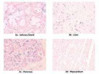
Gross • Encapsulated • Homogeneous, smooth • Orange/rust color • Histology • Cords of uniform cells and thin fibrous stroma Well demarcated/encapsulated
Histopathology • Composed of uniform oncocytic cells arranged in solid, tubular, or trabecular , membranous patterns • The individual cells are polygonal, with characteristically abundant granular cytoplasm and round nuclei with eccentric nucleoli.
Electron microscopy: • Mitochondrial hyperplasia • 60% of cell volume
Monomorphic Adenomas • Basal cell, canalicular, sebaceous, glycogen-rich, clear cell • Basal cell is most common: 1.8% of benign epithelial salivary gland neoplasms • 6th decade • M:F = approximately 1:1 • Caucasian > African American • Most common in parotid
Basal Cell Adenoma Solid • Most common • Solid nests of tumor cells • Uniform, hyperchromatic, round nuclei, indistinct cytoplasm • Peripheral nuclear palisading • Scant stroma
Basal Cell Adenoma • Trabecular • Cells in elongated trabecular pattern • Vascular stroma
Basal Cell Adenoma • Tubular • Multiple duct-like structures • Columnar cell lining • Vascular stroma
Basal Cell Adenoma • Membranous • Thick eosinophilic hyaline membranes surrounding nests of tumor cells • “jigsaw-puzzle” appearance
Monomorphic Adenomas • Canalicular adenoma • 7th decade • F:M – 1.8:1 • Most common in minor salivary glands of the upper lip (74%) • Painless submucosal mass
Canalicular Adenoma • Histology • Well-circumscribed • Multiple foci • Tubular structures line by columnar or cuboidal cells • Vascular stroma
Myoepithelioma • <1% of all salivary neoplasms • 3rd-6th decades • F>M • Minor salivary glands > parotid > submandibular gland • Presentation: asymptomatic mass
Myoepithelioma • Histology • Spindle cell • More common • Parotid • Uniform, central nuclei • Eosinophilic granular or fibrillar cytoplasm • Plasmacytoid cell • Polygonal • Eccentric oval nuclei