
Gait Analysis
E N D
Presentation Transcript
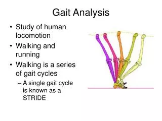
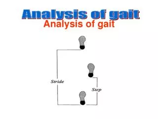
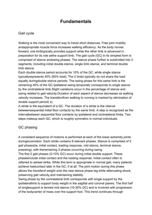
Fundamentals Gait cycle Walking is the most convenient way to travel short distances. Free joint mobility andappropriate muscle force increases walking efficiency. As the body moves forward, one limbtypically provides support while the other limb is advanced in preparation for its role asthe support limb. The gait cycle (GC) in its simplest form is comprised of stance andswing phases. The stance phase further is subdivided into 3 segments, including initial double stance, single limb stance, and terminal double limb stance. Each double stance period accounts for 10% of the GC, while single stance typicallyrepresents 40% (60% total). The 2 limbs typically do not share the load equally duringdouble stance periods. The swing phase for this same limb is the remaining 40% of the GC.Ipsilateral swing temporally corresponds to single stance by the contralateral limb.Slight variations occur in the percentage of stance and swing related to gait velocity.Duration of each aspect of stance decreases as walking velocity increases. The transitionfrom walking to running is marked by elimination of double support period( s). A stride is the equivalent of a GC. The duration of a stride is the interval betweensequential initial floor contacts by the same limb. A step is recognized as the intervalbetween sequential floor contacts by ipsilateral and contralateral limbs. Two steps makeup each GC, which is roughly symmetric in normal individuals. GC phasing A consistent sequence of motions is performed at each of the lower extremity joints duringlocomotion. Each stride contains 8 relevant phases. Stance is comprised of 5 gait phases(ie, initial contact, loading response, mid stance, terminal stance, preswing), with theremaining 3 phases occurring during swing. The first 2 gait phases (0-10% GC) occur during initial double support. These phasesinclude initial contact and the loading response. Initial contact often is referred to asheel strike. While this term is appropriate in normal gait, many patients achieve heelcontact later in the GC, if at all. The joint motion during this phase allows the transferof weight onto the new stance phase leg while attenuating shock, preserving gait velocity,and maintaining stability. Swing phase by the contralateral limb corresponds with single support by the ipsilaterallimb to support body weight in the sagittal and coronal planes. The first half of singlesupport is termed mid stance (10-30% GC) and is involved with progression of the bodycenter of mass over the support foot. This trend continues through
terminal stance (30-50%GC). This phase includes heel rise of the support foot and terminates with contralateralfoot contact. [1] The final stance element, preswing (50-60% GC), is related functionallymore to the swing phase that follows than to the preceding stance phase events. Preswingbegins with terminal double support and ends with toe-off of the ipsilateral limb. Three unique phases characterize swing, including initial swing (60-73% GC), mid swing(73-87% GC), and terminal swing (87-100% GC). The swing phase achieves foot clearance andadvancing of the trailing limb. Shock absorption Shock absorption and energy conservation are important aspects of efficient gait. Alteredjoint motion or absent muscle forces may increase joint reaction (contact) forces and leadsubsequently to additional pathology. In early stance, nearly 60% of one's body weight isloaded abruptly (less than 20 milliseconds) onto the ipsilateral limb. This abrupt impactis attenuated at each of the lower extremity joints. Loading response plantar flexion ispassive, substantially restrained by eccentric work of pretibial muscles. The absorptivework by pretibial muscles delays forefoot contact until late in the initial double supportperiod (7-8% GC). At initial contact, external (ground reaction) forces applied to the contact foot producea tendency toward knee flexion. Repositioning the knee (recurvatum) increases kneemechanical stability, but at the cost of increased contact forces and shock generation. Abalance between knee stability and shock absorption is achieved by eccentric quadricepscontractions during loading response. The impact of loading is minimized at the hip duringsingle support through hip abductor muscle contraction. Energy conservation Ambulation always is associated with metabolic costs. These costs are relatively minor innormal adults performing free speed level walking. The self-selected walking speed innormal adults closely matches the velocity that minimizes metabolic work. This associationdoes not apply with gait pathology. Walking velocity, energy cost per time, and energycost per distance are considerations when the patient is making choices about walkingversus wheelchair mobility. Gait velocity typically decreases with all neuromuscularpathology, and the reduction is related to the magnitude of the pathology. Energy cost perunit of time may not change substantially, even with severe involvement. Energy cost perunit of time is maintained by decreasing walking velocity considerably. Energy cost perunit of time does not change markedly following stroke, as compared to changes associatedwith aging; however, the energy requirement per distance traveled is more than 3 timesnormal.
In this same population, wheelchair use cuts energy cost per distance in half anddecreases cost per minute slightly, while preserving ambulation velocity. Similar trendsare observed when examining various energy cost parameters in individuals with spinal cordinjury, myelomeningocele, and increasing levels of amputation. Energy cost to travel aprescribed distance increases (greater than 500% increase in myelomeningocele withbilateral knee-ankle-foot orthoses), while oxygen cost per minute is maintained bydecreasing walking velocity substantially. Often the critical factor in selecting awheelchair for mobility is the energy requirement to traverse a given distance. Mostindividuals self-select wheelchair mobility when cost per distance exceeds 300% of normalvalues. Methods Of Analysis Most commonly, observational gait analysis is appropriate to characterize most gaitpathologies. This approach is sufficient to note gross abnormalities in walking; however,as walking complexity increases with organic pathology, objective analysis becomesnecessary. The measurement systems that follow may be used individually or collectively.The choice of method is based on clinical need, financial considerations, and staffing atthe specific laboratory. Typically, motion and force data are compiled simultaneously. Motion Instrumented motion analysis is a logical extension of observational gait analysis. Thelevel of complexity involved in examining 3 joints each in 2 limbs is such that fewindividuals can perform this task consistently. Measurement of human motion is complex.Although most gait motion occurs in the sagittal plane, subtle rotations in the otherplanes are clinically important. Analysis of motion in the sagittal plane is confounded ifthat motion does not lie within the plane recorded by a single camera. Rotations of thelimb are attenuated when the limb is internally or externally rotated with respect to thecamera's plane. Three-dimensional motion analysis overcomes this drawback. Position of joint centers of rotation is estimated by markers placed on the skin surface.Markers may be enhanced with passive markers or active light-emitting diodes. Motion ofthe limb segments about the joint centers is recorded. At least 3 markers are required perlimb segment, and the position of each marker is recorded using multiple cameras. Acalibration system is used to translate film to real-life dimensions. Angular position ofeach segment may be determined for each percent GC interval. Angular velocity andacceleration of the limb segments are obtained by mathematical differentiation andsmoothing of limb position data. Motion data may be combined with analysis of externalforces that act on the body.
An alternative technique to quantify motion is through use of electrogoniometers. Atriaxial electrogoniometer has parallelograms and potentiometers for the sagittal,coronal, and transverse planes. Electrogoniometers have several limitations, but theadvantages of this method include convenience, ease of use, immediate availability ofdata, and decreased expense when compared to video acquisition techniques. External forces Calculation of joint moments (torque) and reaction forces between segments is dependent onknowledge of the inertial components of the respective segments (kinematics), bodysegment parameters, and external forces (kinetics) that affect the body. The magnitude and distribution of segment masses with respect to joint axes are obtainedfrom cadaveric data or mathematical modeling of limb segments. The contact (groundreaction) force applied at the distal segment is measured with a force platform. This thinplate typically measures forces and moments in 3 dimensions about the foot center ofpressure. Oscillation of the center of pressure may be obtained in the same manner. Moments and power Integration of external force, center of pressure, unique body segment parameters, andmotion data yields information on joint moments, joint power, and reaction forces betweensegments using standard inverse dynamics techniques. The role of muscle groups is inferredfrom the magnitude and sign of the moments and power at the respective lower extremityjoints. Dynamic electromyography Muscle action can not be measured directly. The electromyogram (EMG) allows indirectmeasurement of muscle activity. While single motor units are analyzed routinely inclinical electrodiagnostic studies, this level of analysis is not performed as part of aroutine gait analysis. A typical kinesiologic (dynamic) EMG represents the activity ofmultiple motor units. The stochastic nature of the waveform confounds simple analysis. Theinterference pattern recorded demonstrates both mechanisms used to increase force outputand increase in the number and in the firing rate of motor units. Kinesiologic EMG contains important information about the timing and relative intensity ofthe signal. Timing of the signal is straightforward. Muscle electrical activity precedesforce generation by 40-120 milliseconds. This electromechanical delay is
associated withcompliance of the tendon at the onset of the EMG signal and continued binding of actin-myosin crossbridges after termination of neural drive related to delays in calciumresequestration. Muscle force can not be estimated directly from the relative intensity of the signal. Alinear relationship between EMG intensity and force output has been demonstrated only inisometric contractions. Presentation of the quantified EMG may be in absolute values(volts) or as a percentage of some normalized standard. Absolute voltage does notrepresent clinically significant effort, as the electrode type and placement substantiallyaffect signal magnitude. Normalization of muscle force output allows for comparisonsacross muscles related to relative intensity. Gait EMG is expressed as a percentage ofmaximum muscle contraction. Another technique is to express gait EMG output as apercentage of gait maximum. This latter technique often must be used in persons who cannot give a maximum voluntary contraction as a result of decreased neural control. Adisadvantage is that peak values of weak and strong muscle forces are defined as 100%. Mechanical and metabolic efficiency Mechanical work is the integral of force and velocity over time and the product of jointpower and joint angular velocity. Mechanical work includes changes in the mechanicalenergy of the body links; energy expenditure for mechanical movement as the sum of thepositive values, including general center of mass energy changes (external work); and changes in energy in the links in their movement relative to the center of mass (internalwork). Limb movement requires energy for muscle contraction. Mechanical work does not equalmetabolic work in most instances. For example, mechanical work in a cyclic motion withconstant average speed (without dissipation of energy) is equal to 0 since the mechanicalenergy of the system has not changed (net 0 paradox). Metabolic energy is required formechanical movement in this case. Energy metabolism is another means of assessing the costof locomotion. Energy expenditure may be determined by indirect calorimetry; however, in most recentliterature, the O2 gas volume is reported without conversion to calories. The volume ofoxygen consumption per body weight enables intersubject comparisons and providesinformation on overall gait performance. Measurement of oxygen consumption (metabolicenergy expenditure, VO2) generally is reported under standard conditions of temperature (0° C), pressure (760 mm Hg), and dry conditions (no water vapor). After 2-3 minutes ofexercise at a constant submaximal workload, the rate of oxygen consumption reaches a levelsufficient to meet tissue demand for oxygen. Parameters of physical work (cardiac output,heart rate, and respiratory rate) reach a steady state. Energy consumption for theactivity at this time is reflected by
the rate of oxygen consumption. The O2 ratedetermines the intensity and duration the exercise can be performed. The rate of oxygen consumption is lowest at comfortable walking speeds in normal adults(approximately one third of maximal aerobic capacity). Slight increases in energy demandsare demonstrated at slower walking speeds; more substantial increases are observed aswalking speed increases above the upper limits of normal. Energy expenditure at eachwalking speed is not related to whether the patient is male or female. Ankle/Foot The arcs of motion at the ankle are relatively small; yet, they are essential for shockabsorption and progression of the body's center of mass. The ankle plantar flexesthroughout loading response. Dorsiflexion begins with single support, as the tibia rotatesforward over the fixed foot. Rapid plantar flexion begins at terminal double support, withmaximum plantar flexor position of 30 ° attained at toe-off. This action marks theinitiation of swing with dorsiflexion throughout the 3 swing-phase segments. Motor control at the ankle is understood most easily by commencing with swing phase muscleactivity. Ankle dorsiflexors undergo brief eccentric contraction in preswing, followedshortly by concentric contraction at the initiation of swing. This contraction sequenceincreases mechanical efficiency and assures foot clearance. Continued pretibial muscleisometric contraction through swing maintains this neutral or slightly dorsiflexedposture. Subsequent loading response is notable for eccentric contraction of the pretibialmuscles to control plantar flexion. A brief period of co-contraction of ankle plantarflexors and dorsiflexors occurs at the transition from initial stance double support tosingle support. This co-contraction interval increases limb stability and may smooth thetransition from double to single support. The orientation of the ankle and subtalar axes couples loading response dorsiflexion witheversion. Both are attenuated by eccentric contraction of the ankle inverters (ie,posterior tibialis). The plantar flexors begin force generation in single support, withpeak activity late in terminal stance and preswing. The absorptive power demonstrated insingle support is related to their role in restraining forward tibial rotation. The timingof the triceps surae and perimalleolar muscles is similar to that of the triceps surae(gastrocnemius plus soleus) that supplies most of the ankle plantar flexor moment. Theankle then is plantar flexed vigorously during preswing. The role of the calf muscles during preswing is controversial. Some authors describe thisgait phase as push-off and suggest that muscle force by the ipsilateral calf activelypropels the limb (and body center of mass) forward. An alternative model is proposed byPerry, who states that the ipsilateral limb makes no active contribution to the push-off.A third model is that active plantar flexion occurs at the ankle (positive
power) and doesnot serve to propel the body forward (no increase in walking velocity), but, instead,functions as part of a closed kinetic chain to initiate knee and hip flexion inpreparation for swing. This phenomenon may account for the increased knee and hip powerrequirements in individuals who do not plantar flex actively in preswing (ie, transtibialamputees). Knee Most knee motion is limited to the sagittal plane. The knee travels from slight kneeflexion at initial contact (5 ° )to nearly 20 ° of flexion by the end of loading response.The knee then extends( net flexion )through single support, with peak stance phaseextension at 40% GC. At the conclusion of terminal stance and preswing, knee flexion israpid, continuing through initial swing until peak knee flexion (60 ° )occurs. This trendthen is reversed, with knee extension continuing through terminal swing. Peak kneeextension occurs slightly before the end of the swing phase, with minor flexion occurringin preparation for the subsequent stance phase. The role of knee muscles, like that of ankle muscles, is understood most easily if onebegins the analysis as the limb is in swing. In early swing phase, knee flexion ispassive, resulting from active plantar flexion and hip flexion. During swing, motion atthe hip changes from flexion to extension, with the knee passively extended as a result ofthis change of hip joint direction. All 3 hamstrings (long head of the biceps femoris,semimembranosus, semitendinosus) are active in mid-to-late swing to decelerate theextending knee. The quadriceps is also active in terminal swing. This co-contraction ofknee flexors and extensors prepares the limb for weight acceptance that follows shortly.The quadriceps continues to be active in loading support. The eccentric activity of thesemuscles attenuates the shock of weight acceptance, while preventing excess knee flexion (<20 °). Both the hamstring and quadriceps muscles are quiescent during mid stance whilethe knee is extended passively. The role of the remaining quadriceps muscle is different.The rectus femoris is active electrically for only a brief period corresponding withpreswing and initial swing. Although this muscle crosses and extends the knee, its role ingait is to assist in flexion at the hip as swing commences. Hip The gluteus maximus timing and relative intensity are similar to those demonstrated by theother hip extensors (eg, hamstrings). During late swing, the gluteus maximus functions toreverse hip flexion to extension. The gluteus maximus, generally the body's singlestrongest muscle, resists external forces from loading response that would flex the hipexcessively (eg, jack-knifing). A similar EMG profile and role are demonstrated by theadductor magnus. Unlike in the knee and ankle, there is a
substantial amount of frontalplane motion at the hip. During single support, the mass of the torso tends to rotate thebody about the stance limb hip joint axis (eg, contralateral lateral tilt). Typically,this motion is limited by eccentric activity of the gluteus medius, gluteus minimus, and,to a lesser extent, the tensor fascia lata. Weakness Insufficient force output by muscles may result from disuse, primary muscle disease, orneurologic impairment. Substitution of impaired motor function frequently is possible, andreduction of walking velocity further reduces demand on muscles. Muscle force requirementsare trivial in normal individuals. Less than 25% of normal strength (manual muscle testgrade 3+/ 5) is needed for locomotion. In cases of disease, with decreased maximum muscleforce, functional reserve is lost. Endurance is compromised, and community-levelambulation may not be possible. Selective loss of muscles (or muscle groups) isdemonstrated by characteristic gait patterns. In each case, orthotic management istargeted at preventing excessive motion. Uncompensated calf weakness results in diminished midstance control of the forwardlyrotating tibia. Substitution for calf weakness is difficult. The increased amount and rateof dorsiflexion increases the functional demands on the quadriceps to maintain limbstability. Alternatively, tibial advancement is curtailed with resultant genu recurvatum,decreasing the work of the quadriceps. Unfortunately, this gait alteration frequentlyinitiates knee joint pathology. Other compensatory maneuvers include reducing stepvelocity and step length. The consequence of pretibial muscle weakness is increased plantar flexion. With mildweakness, foot slap occurs during loading response. With more extensive dorsiflexionweakness, foot drop and toe drag are observed during swing phase. Compensation is achievedthrough circumduction of the ipsilateral limb, increased ipsilateral knee plus hipflexion, and hip hiking of the contralateral limb. Each of these tendencies assists inswing-phase toe clearance. Of these compensations, circumduction is the most energyefficient and most commonly observed technique for dorsiflexor insufficiency. Quadriceps weakness diminishes knee control, and deficits in stance are most pronounced.External forces tend to hyperflex the knee, and a variety of compensations are employed topreserve stability in weight bearing. Compensations begin prior to weight acceptance (lateswing) and continue through that limb's support activity. Late swing hip flexion leads topassive knee extension through momentum transfer. Stance phase increased knee flexion isattenuated by hip extension and premature plantar flexion. Alternatively, externalrotation of the affected limb orients the external force vector medial to the joint axis,minimizing the tendency toward knee flexion.
Isolated hamstring weakness is an uncommon gait deficit. Isolated hip flexor weakness isuncommon. Clinical presentation includes decreased walking velocity or decreased hip rangeof motion. Reduction of hip extensor forces, which may be secondary to paralysis or musclelengthening, leads to increases in anterior pelvic tilt during stance. Hip adductor weakness results in pelvic instability during stance. External forces (ie,torso mass) lead to frontal plane rotation about the ipsilateral hip joint. Thiscontralateral tilt normally is resisted by eccentric contraction of ipsilateral hipabductors (ie, gluteus medius, minimus). Shifting the torso over the ipsilateral hip jointin mid stance (Trendelenburg gait) minimizes the force required by the hip abductors. Thisgait pattern, which limits contact forces between femur and acetabulum, also is observedin hip joint pathology. Spasticity Spasticity results from lesions of the CNS. The primary lesion may be in the brain orspinal cord, although multiple lesions along the neuraxis also are possible. Spasticitymay involve the upper and lower extremities. Although arm swing does have some effect ongait and is altered with spasticity, discussion of gait alterations resulting from upperextremity spasticity is beyond the scope of this analysis. In the lower extremity, a fewcommon patterns emerge, including the equinovarus foot, valgus foot, striatal toe, stiff(extended) knee, flexed knee, adducted thighs, and flexed hip. Equinovarus is the most common spasticity-related pathology seen in the lower extremity.Toe curling (clawing) may coexist. Contact is at the forefoot, with weight acceptancealong the lateral aspect of the foot. Skin breakdown along the lateral border of the footmay be an associated problem. Equinovarus posturing limits single support dorsiflexion, prevents forward progression ofthe tibia, and leads to a hyperextension thrust of the knee and restrained forwardtranslation of the body's center of mass. This abnormality compromises stability bydecreasing the base of support and may prevent functional ambulation. Swing phase footclearance is compromised. Muscles that may contribute to this disorder include tibialisposterior, tibialis anterior, long toe flexors, medial and lateral gastrocnemius, soleus,extensor hallucis longus, and peroneus longus. Conservative management includesadministration of neurolytic and chemodenervation agents. Tendon lengthening decreasesforce output with no change in neural drive. Valgus foot results from inappropriate contraction of peroneal and triceps surae muscles.Stance phase stability is compromised by an abnormal base of support. This condition alsomay lead to genu valgum stresses and resultant pathology to medial knee joint structures.
Persistent great toe hyperextension (hitchhiker toe) is not uncommon. Selection offootwear is complicated. The usual offender is the extensor hallucis longus (EHL) muscle.This pathology often is managed successfully with motor point block of the EHL muscle.Diagnostic blocks often are helpful if co-contraction by the flexor hallucis longus issuggested. Stiff knee, a frequent complication of stroke, is a severe hindrance during swing.Overactivity by iliopsoas, gluteus maximus, quadriceps, and hamstrings (as hip extensors)is a possible contributor to this condition. Functional deficits include increasedeffective limb length, with circumduction, contralateral vaulting, or hiking of the pelvisrequired for foot clearance. Energy consumption is increased, typically in patients withlittle functional reserve. Kinesiologic EMG analysis may be required to identify thecontributing muscle( s) from the extensive aforementioned list. Crouched gait is marked by excessive knee flexion in both swing and stance. Limbadvancement is attenuated as part of this crouch pattern. Both hamstring and quadricepsmuscles may be involved. Kinesiologic EMG may demonstrate prolonged activity of the medialhamstrings. Knee flexion contracture is common. Excessive hip flexion may interfere withmultiple activities of daily living and may contribute to knee flexion deformity. Again,excessive hip flexion may interfere with limb advancement in swing, as well as decreasedadvancement of the center of mass over the flexed support limb. Initial treatment of bothexcessive knee and hip flexion is controversial. Therapeutic interventions include motorpoint blocks, chemoneurolysis, tendon lengthening, tendon transfers, and muscle release. Thigh adduction (ie, scissoring) may interfere with daily activities, as well as withgait. Limb advancement is curtailed by severe hip adduction. Balance may be impaired as aresult of a narrowing of the base of support. Muscles that potentially contribute to gaitscissoring include the adductor longus and brevis, adductor magnus, and gracilis. Lessfrequently, the iliopsoas and pectineus are contributors. Diagnostic obturator nerve blockhelps establish whether contracture is present. A study by MacWilliams et al found that among patients who underwent a hamstring-lengthening procedure, those whose surgeon followed or agreed with a gait analysis--derived surgical recommendation had a 3.6-fold greater chance of a positive outcome. Contractures Plantar flexor contracture of 15 ° is most common. This joint position minimizes jointcapsule tensile forces. Contracture may be rigid or elastic. Only the ankle orientation atinitial contact and mid swing is altered with elastic contractures. Despite that fact,limb advancement may be slowed, because of the viscoelastic nature of tissues. A rigid 15° contracture is associated with early foot flat and lack of early stance phase tibialadvancement. Decreased toe clearance and foot drag are swing
phase consequences.Dorsiflexion contractures are rare, although sometimes they are observed in lumbar-levelmyelomeningocele (present anterior tibialis and absent triceps surae). Knee joint contractures are typically at 30 ° of flexion. This position correlates withthe resting position of the swollen knee. Only initial swing is affected by thisposturing. Step length is shortened as a result of decreased knee extension in terminalswing, and metabolic demands are increased. Knee extension contractures (ie, capsulescarring) diminish limb advancement in early swing phase. Hip flexor contractures result from shortening of hip flexor muscles or the joint capsule.A tight iliotibial band is a variant of this disorder. Each results in increased forwardpelvic tilt. Loss of hip joint flexion due to hip extensor contracture is infrequent. Pain Pain does not inhibit normal motion directly; however, deformity and weakness may resultfrom the person's attempts to attenuate pain through gait modifications. The positions ofmaximal joint capsule and ligamentous laxity typically are adopted. Generally, jointreaction forces are magnified with increases in muscle forces crossing the joint, andincreased joint contact force is associated with increased discomfort. One mechanism forreduction of joint pain is to limit the muscle force output at the painful joint. Thisprotective response leads to muscle atrophy and weakness. Sensory loss Impaired proprioception inhibits walking because of diminished information about the limbsegment positions in space. When sensory loss predominates (intact motor control), initialcontact may be emphasized. Superimposed motor losses diminish the individual's ability tomake rapid substitution efforts, resulting in decrements in walking velocity andstability. Orthotics and assistive devices Studies have indicated that when required, an ankle-foot orthosis (AFO) and/or crutchesmay improve energy requirements and some kinematic properties of gait in children withspina bifida. https://nydnrehab.com/treatment-methods/gaitanalysis/