Download
1 / 8
80 likes | 122 Vues
Direct Plagiarism

E N D
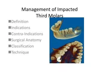
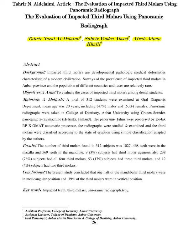
Tahrir N. Aldelaimi Article : The Evaluation of Impacted Third Molars Using Panoramic Radiograph The Evaluation of Impacted Third Molars Using Panoramic The Evaluation of Impacted Third Molars Using Panoramic Radiograph Radiograph Tahrir Nazal Al-Delaimi1 , Suheir Wadea Abood2, Afrah Adnan Khalil3 Abstract Background:Impacted third molars are developmental pathologic medical deformities characteristic of a modern civilization.Surveys of the prevalence of impacted third molars in Anbar province and the population of different countries and races are relatively rare. Objectives & Aims: To evaluate the cases of impacted third molars among dental students. Materials & Methods: A total of 312 students were examined at Oral Diagnosis Department, mean age was 20 years, including (47%) males and (53%) females. Panoramic radiographs were taken in College of Dentistry, Anbar University using Cranex-Soredex panoramic x-ray machine (Helsinki, Finland). The panoramic Films were processed by Kodak RP X-OMAT automatic processor, the radiographs were studied & examined and the third molars were classified according to the state of eruption using simple classification adapted by the authors. Results: The number of third molars found in 312 subjects was 1027; 468 teeth were in the maxilla and 569 teeth in the mandible. 9 (3%) subjects had third molar agenesis also 238 (76%) subjects had all four third molars, 53 (17%) subjects had three third molars, and 12 (4%) subjects had two third molars. Conclusions: The present study concluded that one half of the mandibular third molars were in mesioangular position and 39% of the third molars were in vertical position. Key words: Impacted teeth, third molars, panoramic radiograph,Iraq. 1Assistant Professor, College of Dentistry, Anbar University. 2 Assistant Lecturer, College of Dentistry, Anbar University. 3Oral Pathologist, Anbar Health Directorate & College of Dentistry, Anbar University. 26
Tahrir N. Aldelaimi Article : The Evaluation of Impacted Third Molars Using Panoramic Radiograph Introduction Surveys of the prevalence of impacted third molars in the population of different countries and races are relatively rare. Mead 1 reported that 18.8% of his office patients had at least one impacted tooth. Hellman 2 who examined 433 students University found an incidence of 15.2%, with females twice as likely to have an impaction as males. Dachi and Howell 3 examined 3874 patients and found that 29.9% of the maxillary third molar and 17.5% of the mandibular third molar were impacted but found no sex difference. Kramer and Williams 4 surveyed a black population and found the incidence of impaction was 18.2%. They also found that third molars were more often impacted in the maxilla than in the mandible, but there was no sex difference. Studies in Scandinavian communities revealed that the proportion of persons with impaction of third molar varies from 19 to 35% 5,6,7,8. Agenesis of one or more third molars vary substantially in persons from different races, with prevalence of approximately 1% in African Negro and Australian aboriginal samples 14, 10% to 25% in whites 9,10,11,12,14,15, 19% to 35% in Scandinavian 8,16 and 30% in Japanese 17 and Chinese 18. Levesque et al.19 reported 9% bilateral agenesis of mandibular third molar, without significant distribution. Gorgani et al. 12 found that the rate of agenesis of third molars for black and white population ranged from 7% to 10% with bilateral agenesis occurring in 79% of the sample. Times of eruption of third molars are also variable starting at the age of 16 years; averages of 17.8 10, 19.9 19, 20.5 20, and 24 eruption of third molars was reported among rural Nigerians, that is, by age of 13 years for females and 15 years for males 22. In most of these studies, however, the criteria used for eruption were the emergence of any portion of the crown through the oral mucosa. This may give misleading results because many of the third molar do not continue to erupt but remain impacted in a partially erupted position 23. at Columbia Material and Methods A total of 312 dental students were seen in the department of Oral Diagnosis, College of Dentistry, Anbar University. Subjects receiving orthodontic treatment were excluded from the survey as well as those who had any of the posterior teeth, excluding third molars, extracted. The mean age was 20 years, including 148 (47%) males and 164 (53%) females. Panoramic radiographs were taken in college of Dentistry, Anbar University using Cranex-Soredex panoramic x-ray machine (Helsinki, Finland). The panoramic Films were processed by Kodak RP X-OMAT automatic processor, the radiographs were studied & examined and the third molars were classified according to the state of eruption using simple classification adapted by the authors that classified the third molars according to the state of eruption in two categories: (1) erupted (partly or completely erupted), the highest portion of the tooth was above the cervical line of adjacent second molar; (2) nonerupted (bony or soft tissue impaction) but with complete root formation. Impacted third molars were also grouped according to their position including vertical, mesioangular, distoangular, and horizontal. Nonerupted third molars with incomplete root formation were not included in the group of impacted teeth because eventual eruption is uncertain; also authors advocate a simple classification that group maxillary and mandibular third molars according to depth by their relationship to the cervical line of adjacent second molars. difference in sex 21years have been reported. Earlier 27 Vol.8,No.1,August 2010 , ISSN: 2070-8882
Tahrir N. Aldelaimi Article : The Evaluation of Impacted Third Molars Using Panoramic Radiograph In level A, the highest part of the third molars was on the same level or above the occlusal plane of adjacent second molars. In level B, the highest part of the third molar was below the occlusal plane but above the cervical line of the second molar. In level C, the highest part of the third molar was beneath the cervical line of the second molar. All erupted and impacted third molars were registered except the unerupted third molars with incomplete root formation. The maxillary and mandibular nonerupted third molars were classified according to the stage of root formation, namely, noncompleted. The noncompleted were divided into one third or two third complete formations. Results The number of third molars per person is presented in Table I. The number of third molars found in 312 subjects was 1027; 468 teeth were in the maxilla and 569 teeth in the mandible. 9 (3%) subjects had third molar agenesis also 238 (76%) subjects had all four third molars, 53 (17%) subjects had three third molars, and 12 (4%) subjects had two third molars. The angular position of third molars presented in table II which showed that of the total 1027 teeth; 32% were vertical positioned, 57% were mesioangular, 7% distoangular and 4% were horizontally positioned. completed or Table I. Number of third molars per person Number of third molars Sex Total Four Three Two One None Male 88 28 22 6 4 148 (47%) Female 107 25 21 6 5 164 (53%) Total 195 53 43 12 9 312 (100%) The level of eruption of third molars is shown in Table III; of the 688 teeth, 399 (58%) were positioned with their occlusal surfaces on the same level or above the occlusal plane of the adjacent second molars (level A). Females demonstrate a higher frequency (9%) of level A eruption than males. The maxilla is predominant site (226 teeth) over the mandible (173 teeth). The difference was statistically significant ( p < 0.05, x2 = 7.03). Level B eruption was the least frequent in occurrence among the other levels of eruption ( Table III). Vol.8,No.1,August 2010 , ISSN: 2070-8882 28
Tahrir N. Aldelaimi Article : The Evaluation of Impacted Third Molars Using Panoramic Radiograph Females had 4% more third molars at level B eruption than males. Level B eruption showed a higher frequency in the maxilla (61 teeth) than in the mandible (46 teeth). The difference was not statistically significant. One hundred eighty third molars (26.2%) were erupted to level C. The proportion of level C eruption in males was higher (13.1%) than in females. The difference was highly significant ( p < 0.001, x2 test ). Level C eruption occurred more frequently in the maxilla than in the mandible at a ratio of 1.3 ; However, the difference was not statistically significant. The development of roots was assessed in unerupted mandibular third molars. Of 232 subjects, 27 (11.6%) had incomplete root formation. This corresponds to 8.4% (68 of 814) of the total number of teeth examined. The ratio of two thirds to one third root formation was 4.3. One fourth of the unerupted third molars showed incomplete root formation. There was a tendency for more frequent incomplete root formation in males than in females. maxillary and Table II. Percent of angular position of mandibular third molars Angular position Total Vertical Mesioangular Distoangular Horizontal 329 (32%) 585 (57%) 72 (7%) 41 (4%) 1027 Table III. Level of eruption of maxillary and mandibular third molars Level of eruption Sex Total A* B** C*** Male 165 (53%) 43 (14%) 104 (33%) 312 Female 234 (62%) 66 (18%) 76 (20%) 376 Total 399 (58%) 109 (16%) 180 (26%) 688 * A, the highest part of the third molars was on the same level or above the occlusal plane of adjacent second molars. ** B, the highest part of the third molar was below the occlusal plane but above the cervical line of the second molar. *** C, the highest part of the third molar was beneath the cervical line of the second molar. Vol.8,No.1,August 2010 , ISSN: 2070-8882 29
Tahrir N. Aldelaimi Article : The Evaluation of Impacted Third Molars Using Panoramic Radiograph Discussion Third molars are the teeth that are most often congenitally missing. If present, third molars may follow an abortive eruption path and become impacted as a result of the skeletal insufficiency in the area where they normally erupt. Impacted third molars are developmental pathologic deformities characteristic of a modern civilization. They account for 98% of all impacted teeth 24. The cause of agenesis of one or more teeth is essentially unknown, but several mechanisms have been suggested: physical disruption of the dental lamina, space limitation, and an inherent defect of the dental lamina or failure of induction of the underlying mesenchyme. Space limitation and crowding is particularly involved in the agenesis of third molars, where competition for minimum nutritional requirement constricted area can cause tooth germ regression and agenesis14. In general, these changes are under the influence of genetic and environmental factors. There is no known association of agenesis of only the third molars with syndromes. On the other hand, impaction of third molars occurs as a result of retardation of facial growth, shortage of space in the third molars region, vertical direction of the condylar growth associated with low resorption of the anterior border of the ramus, the distal direction of the eruption of the other teeth, low mandibular growth rate resulting in a reduction in the length of the jaws, early physical maturity, and late third molars mineralization 5,25,26,27. The growth of the maxilla and mandible is essentially completed by 16 to 17 years of age. The mean age of participants in the present study (20.4 years) is very close to the average age of 20.3 years reported for the eruption of third molars 2,11,19,21. Schersten et al.8 suggested that 20 to 25 years is the most suitable age for studying the frequency of third molars and its impaction. The reason for this is to avoid overestimation of third molars agenesis as a result of unnoticed early extraction in older group. Further, many impacted third molars can change their position and erupt after the age of 18 to 20 years 23,28,29,30. This indicates that the eruption period for third molars is longer than supposed previously. Results of the present study showed that about three quarters of the subjects had all four third molars. This proportion is higher than Hellman 2 found with American students and Schersten et al.8 with Scandinavians who also noted that one half of the persons had all four third molars. Our observation that 9.1% of third molars are congenitally missing is in agreement with the data reported by Levesque et al.19 for French-Canadians, Gorgani et al.12 for American whites, and Haider and Shalhoub 31 for the Saudi population but one half the prevalence reported by Porgel 32 for orthodontic patients. In the present study, 1.7% had agenesis of all third molars. This observation is considerably less than that for the Scandanavian population (10% to 13%) 5,8,16and the Americans (7% to 10%) 12. According to Banks 9, it is most common for two third molars to be missing, followed by one, four, and three. On the contrary, we found the order of frequency for missing teeth is one, two, three, and four, which is in agreement with frequency noted by Nanda 33. The proportion of third molars agenesis for females was less than males, but the difference, unlike the sample of Thompson et al. 15 was not significant. In this context, our findings are in agreement with those reported by Levesque et al.19 and Gorgani et al.12 but differ from those of Hellman 2 and Shah et al. 34 who found that females had a higher incidence of third molars agenesis. medical in a spatially 30 Vol.8,No.1,August 2010 , ISSN: 2070-8882
Tahrir N. Aldelaimi Article : The Evaluation of Impacted Third Molars Using Panoramic Radiograph Results of the present study agree with earlier reports 1,3,4,16 that maxillary third molars were more commonly missing than mandibular third molars. Equal distribution between the left and right side as noted by Hellman 2, Grahen 6, and Shah et al.34 is confirmed in our study. In the present study, one third of the persons had one more impacted third molars. This figure is considerably higher than that reported by Shah et al.34 (6.9%) for Canadian population and about 13% higher than the average of 20% presented by some studies in the United States1,2,3,4, but in Scandinavian data (29% to 35%) 5,7,9. Our results showed no sex differences in the prevalence of third molars impaction. This differs from the observation made by Hellman[2] and Schersten et al.8 but is in agreement with the observation made by Dachi and Howell Williams predominance in the third molars impaction raised the question against Hellman’s statement that the jaws of females stop growing when third molars just begin to erupt, whereas, in males, the growth of the jaws continues beyond the time of eruption of the third molars. Our observation that the maxilla showed a greater tendency for occurrence of third molars impaction than the mandible (52.6% versus 47.4%) is in the general line, but less in magnitude, than those reported by others 2-4. The angulation of the third molars may influence their subsequent eruption and therefore impart clinical significance on the state of third molars 35,37,38. Altonen et al.27 found that the angulation of the lower third molars in relation to second molars decreased with age especially after 14 to 15 years of age. Richardson25 reported that third molars with a small degree of angulation erupted earlier than those with Longitudinal studies on the positional changes and eruption of third molars demonstrate that many unerupted, partially erupted, or impacted third molars are likely to change their position and erupt after the age of 20 years and that their final state remains unpredictable 23,28,29,30. It has been speculated that the third molars have a constant path of eruption until contact is made with adjacent teeth. The present study showed that one half of the mandibular third molars were in mesioangular position (Table II). This number is considerably higher (18%) than that reported by Sewerin and von Wowern 28, but similar in proportion to that noted by Richardson 30 for persons aged 21 years. Our observation that 39% of the third molars were in vertical position is less than that reported by others 29,30. agreement with References 1-Mead SV. Incidence of impacted teeth. INT JORTHOD 1930 ; 16:85-90. 2-llman M. Our third molar teeth: their eruption, presence and absence. Dental Cosmos 1936; 78:750-62. 3-Dachi SF, Howell FV. A survey of 3874 routine full-mouth radiographs :II. a study of impacted teeth. J Oral maxillofac Surg 1961;14:1165-9. 4-Kramer RM, Williams, AC. The incidence of impacted teeth. ORAL SURG ORAL MED ORAL PATHOL 1970 ; 29:237-41. 5-Bjork A, Jensen E, Palling M. Mandibular growth and third molar impaction. ACTA ODONTOL SCAND 1956 ; 14:231-72. 6-Aitasalo K, Lehtenin R, Oksala E. An orthopantomorphic study of prevalence of impacted teeth. INT JORAL SURG 1972 ; 1:117-20. 7-Ylipaavalniemi P, Turtola L, Murtomaa H, Rytomaa I. Evaluation of the need for third molar removals among 20- to 21- years old Finnish university students. PROC FINN DENT SOC 1985;81:222-5. 3 and Kramer and 4. The lack of definite sex steeper angulation. 31 Vol.8,No.1,August 2010 , ISSN: 2070-8882
Tahrir N. Aldelaimi Article : The Evaluation of Impacted Third Molars Using Panoramic Radiograph 8-Schersten E, Lysell L, Rohlin M. Prevalence of third molars in dental students. SWED DENT J 1989;13:7-13. 9-Banks HV. Incidence of third molar development. 1934;4:223-33 10-Adamson K. The controversy concerning the first permanent molar. AUST DENT J 1962;7:191-201. 11-Garn SM, Lewis A, Bonne B. Third molar formation and its development course. ANGLE ORTHOD 1962;32:270-9 12-Gorgani N, Sullivan RE, Dubois L. A radiographic investigation of third-molar development. J Dent Child 1990;57:106- 10. 13-Richardson M. Late third molar agenesis : its significance in orthodontic treatment. ANGLE ORTHOD 1980;50:121-8. 14-Stewart RE, Barber TK, Thoutman KC, Wei SHY. Pediatric dentistry : scientific foundation and clinical practice. 1st ed. St. Louis : CV Mosby, 1982:91. 15-Thompson GW, Popovitch F, Anderson DL. Third molar agenesis in Burlington growth center in Toronto. COMMUNITY DENTAL ORAL EPIDEMIOL 1974;2:187-92. 16-Grahnen H. Hypodontia in the permanent dentition. ODONT REVY 1956:7(suppl):1- 100. 17-Arita M, Iwagak H. Studies on the serial observation of dentofacial region in Japanese children. Monographs. Tokyo : Nikon University School of Dentistry, 1963. 18-Montelius GA. Impacted Teeth : a comparative study of Chinese and Caucasian dentitions. 1932;12:931-8. 19-Levesque GY, Demerjian A, Tanguay R. Sexual dimorphism in the development, emergence, and mandibular third molar.JDENT RES 1981;60:1735-41. 20-Fannig EA. Third molar in Bostonians. AM JPHYS ANTHROPOL 1962;20:151-4. 21-Haralabakis H. Observations on the time of eruption, congenital absence, and impaction of the third molar teeth. TRANS EUR ORTHOD SOC 1957:308-9. 22-Odusanya SA, Abayomi IO. Third molar eruption among rural Nigerians. ORAL SURG ORAL MED 1991;71:51-4. 23-Shiller WR. Positional changes in mesio- angular impacted mandibular third molars during a year. JAM DENT ASSOC 1979;99:460-4. 24-Alling CC, Helfrick JF, Alling RD. Impacted teeth. Philadelphia : WB Saunders, 1993:2,64. 25-Richardson ME. The etiology and prediction of mandibular third molar impaction. ANGLE ORTHOD 1977;47:165- 72. 26-Bishara SE, Andresson G. Third molars : a review. AM JORTHOD 1983;83:131-7. 27-Altonen M, Haavikko K. Mattila K. Developmental position of lower third molar in relation to gonial angle and lower second molar. 1977;47:249-55. 28-Sewerin I, von radiographic four-year follow-up study of asymptomatic mandibular third molars in young adults. INT DENT J 1990;40:24-30. 29-Venta I, Murtomaa H, Turtola L, Meurman J, Ylipaavalniemi P. Clinical follow-up study of third molar eruption from ages 20 to 26 years. ORAL SURG ORAL MED ORAL PATHOL 1991 ; 72:150- 3. 30-Richardson M. Changes in lower third molar position in the young adult. AM J ORTHOD DENTOFACIAL 1992;102:320-7. 31-Haider Z, Shalhoub SY. The incidence of impacted wisdom teeth in a Saudi community. INT ORAL MAXILLOFAC SURG 986;15:569-71. ANGLE ORTHOD ORAL PATHOL ANGLE ORTHOD Wowern N. A JDENT RES ORTHOP agenesis of the 32 Vol.8,No.1,August 2010 , ISSN: 2070-8882
Tahrir N. Aldelaimi Article : The Evaluation of Impacted Third Molars Using Panoramic Radiograph 32-Pogrel H. Radiographic investigation into the incidence of the lower third molar. BR DENT J 1967;122:57-62 33-Nanda RS. Agenesis of the third molar in man. AM JORTHOD 1954;40:698-706. 34-Shah RM, Boyd MA, Vakil TF. Studies of permanent tooth anomalies in 7886 Canadian individuals :II. congenitally missing, supernumerary and peg teeth. J CAN DENT ASSOC 1978;44:265-8. 35-Cryer BS. Third molar eruption and the effect of the lower second permanent molar. DENT PRACT 1967; 16:316-20. 36-Kajii T, Imai T, Kajii S, Iida J. Presence of third molars germs in orthodontic patients in Japan. AJODO 2001;119:245- 50. 37-Shapira J, Chaushu S, Becker A. Prevalence of tooth transposition, third molars agenesis and maxillary canine impaction in individuals with Downs syndrome. The Angle Orthod 2000;70: 290-96. 38-Ghada A Y. Third molar Agenesis. J Bagh Coll Dentistry 2005; 17: 101-104. 33 Vol.8,No.1,August 2010 , ISSN: 2070-8882