
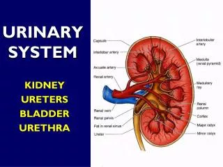
URINARY SYSTEM
URINARY SYSTEM. KIDNEY URETERS BLADDER URETHRA. FUNCTIONS. VOLUME/BLOOD PRESSURE REGULATION Original function Adaptation to terrestrial existence Water and ion retention IONIC BALANCE NITROGENOUS WASTE DISPOSAL A secondarily evolved function Best developed in birds and mammals.

URINARY SYSTEM
E N D
Presentation Transcript
URINARY SYSTEM KIDNEY URETERS BLADDER URETHRA
FUNCTIONS • VOLUME/BLOOD PRESSURE REGULATION • Original function • Adaptation to terrestrial existence • Water and ion retention • IONIC BALANCE • NITROGENOUS WASTE DISPOSAL • A secondarily evolved function • Best developed in birds and mammals
CORTEX & MEDULLA • CORTEX contains mainly tubules; but also RENAL CORPUSCLES • MEDULLA is composed exclusively of tubules • Demarcation line is not sharp
RENAL LOBE • Site of interface with blood vascular system • Blood flow controls action of the nephron • Collecting ducts take thousands of inputs • Depth of lobe & length of loop critical to concentration • Lobe drains at “apex”
THE NEPHRON • Basic functional unit • Millions per kidney • Long and short types • Components: • Renal corpuscle • Proximal tubule • Loop of Henle • Distal tubule • Collecting duct not part of nephron
FUNCTIONAL REGIONS • RENAL CORPUSCLE • Site of initial filtration • PROXIMAL TUBULE • Resorption of water, glucose, proteins and sodium from filtrate: 99% recovery • LOOP OF HENLE • Countercurrent exchange of ions • DISTAL TUBULE • Active transport of sodium • (Collecting duct) • Final concentration of urine
RENAL CORPUSCLE • A URINARY POLE and VASCULAR POLE • GLOMERULUS is the capillary tuft • RENAL CAPSULE has PARIETAL and VISCERAL layers • Capsular space continuous with PCT • Site of initial filtration of urine
RENAL CORPUSCLE • Wall of corpuscle is simple squamous PARIETAL LAYER • Continuous with proximal tubule at urinary pole • Glomerulus covered with specialized VISCERAL LAYER of epithelium, i.e.PODOCYTES
PODOCYTES • From Greek, “podo” = “foot” + “kytos” • Have extensive foot processes to enwrap capillaries of the glomerulus • Thick basal lamina of podocytes is important to filtration • Capillaries are fenestrated
PODOCYTES • Interlaced foot processes cover entire surface of capillary • Large objects retained in lumen (e.g. formed elements); smaller filtered between processes
Blood flow is vital to kidney function EVERYTHING depends on it Primary filtration depends on steady arterial supply and normal pressure Maintenance of normal interstitial osmotic gradient depends on blood flow Specialized arrangements of blood vessel in the kidney serve its needs Main input from renal artery at hilus Routed through arterial branches to individual glomeruli Arcuate Arteries Inter- & Intralobular Arteries Afferent Arterioles Post-glomerular blood flows through a second capillary bed with special function and architecture Drainage of capillary bed is collected and exits via renal vein VASCULAR CONSIDERATIONS
VASCULAR ARRANGEMENTS • Renal Artery supplies main flow • Subdivided into branches • Lobar Arteries • Arcuate Arteries • Smaller branches • Venous return follows reverse path
VASCULAR ARCHITECTURE OF THE CORTEX • Arcuate arteries follow cortico-medullary junction • Give rise to intralobular arteries to feed glomeruli • AFFERENT Arterioles • Rete mirabile arises from EFFERENT Arterioles
AFFERENT & EFFERENT ARTERIOLES • Afferent arterioles arise from intralobulars • Feed glomerular capillary bed • Intralobular arterioles can supply more than one glomerulus • Efferent arterioles feed the capillaries of the RETE MIRABILE
FILTRATION BARRIER COMPONENTS: Glomerular capillary epithelium Basal lamina of capillary Fused basal lamina of podocytes Podocyte processes Basal lamina and SLIT PORE MEMBRANE between podocytes processes Final site of flitration Small molecules (e.g. glucose) pass through; large ones (e.g. proteins > 40 Kd) are retained at various levels Filtration space is continuous with lumen of proximal tubule INITIAL FORMATION OF URINE
PROXIMAL TUBULE • Begins at urinary pole of corpuscle • Initial filtrate essentially blood plasma • Nephron modifies filtrate as it passes • PT is the bulk of the renal cortex • Wall of simple cuboidal epithelium with brush border • Mural epithelial cells extensively interdigitated
PROXIMAL TUBULE • ONLY tubule with a brush border • Often collapsed in LM • Interdigitations obscures boundaries • PINOCYTOSIS for protein absorption
PCT absorbs nearly all the water and glucose in the filtrate • 1.2 liters per MINUTE or more; 99% resorbed • Nearly 1800 liters per day, and 2.5 kg glucose & salts • Protein absorption by engulfment in vesicles at apical surface
LOOP OF HENLE • Loop is the key to formation of a hypertonic urine • Abrupt transition from simple columnar to simple squamous • Thin segments closely applied to blood vessels • “Hairpin turn” at the bottom and return to cortex
LONG & SHORT LOOPS • Cortical nephrons have short loops • Nephrons near medulla have long loops • The longer the loop the more concentrated the urine that nephron can form
DISTAL TUBULE • Begins as ascending portion AFTER loop • Shorter than proximal tubule • No microvilli • Wall simple cuboidal; cells have deep basal folds and numerous mitochondria • Function is active transport of sodium to interstitial space • Special region near corpuscle and afferent arteriole, the MACULA DENSA
MACULA DENSA • Special region of DT • Associated with its OWN corpuscle and arteries that feed its OWN glomerulus • Probably involved in regulation of blood flow • Relationship unclear
JUXTAGLOMERULAR APPARATUS • Macula densa is one component JUXTAGLOMERULAR APPARATUS Other components of the JGA: • Special cells in the wall of the afferent arteriole • Associated cells of uncertain function
JUXTAGLOMERULAR APPARATUS • MD oriented towards afferent arteriole of glomerulus • Special JUXTA-GLOMERULAR CELLS in arteriole wall are secretory • Produce RENIN • “Extra” space filled with mesangial or lacis cells
JGA • COMPONENTS • Macula densa • Juxtaglomerular cells of Afferent arteriole • Renin-secreting • Lacis cells
Renin acts to convert precursor ANGIOTENSINOGEN to ANGIOTENSIN 1 Angiotensin 1 is converted to ANGIOTENSIN 2 Angiotensin 2 causes vasoconstriction & release of ALDOSTERONE from the adrenal gland Net result is retention of water to regulate BP JG cells are part of arterial wall, may be modified from muscle Renin is a HORMONE The kidney is an endocrine organ! Drop in BP triggers release of renin Renin activates the angiotensin system to regulate blood pressure RENIN-ANGIOTENSIN SYSTEM
COLLECTING DUCTS/TUBULES • Key to concentration ability • Accept input from numerous nephrons • Drain out at apex of lobe into pelvis or calyx • Lined with columnar epithelium
POST-GLOMERULAR BLOOD FLOW • The RETE MIRABILE a “wondrous net” • Extensive plexus of capillaries and veins formed AFTER blood has passed out of glomerulus • Surrounds the loop of Henle • Controls Na+gradient of interstitium
RETE MIRABILE & VASA RECTA • Efferent arteriole gives rise to rete • Vasa recta a subdivision • Numerous shunts and cross-links between arterial and venous portions • Recurrent loops and flow parallel and counter to urine flow
CONCENTRATION • Concentration of urine depends on the establishment of a gradient of concentration from “top” to “bottom” • Deep medullary areas have higher osmotic pressure than cortex • The DT provides the ions • The rete and its vasa recta control their distribution • The state of the collecting duct determines final urine concentration
POST FILTRATION EVENTS • Gradient is established in interstitium • First filtrate is passed to the PT and down through gradient • Filtrate loses water by osmosis to increasingly high-osmotic pressure interstitium • Reaches maximum concentration at bottom of loop
LOOP & RETURN • Forming urine rounds the turn and comes back up to cortex • Concentration DECREASES but modification of content is advanced • Distal tubule is actively pumping sodium • Rete is maintaining gradient
ROLE OF COLLECTING DUCT • Urine passes into COLLECTING DUCT for final run down through the gradient • Walls of CD are permeable: water is drawn out and urine is concentrated • COUNTERCURRENT FLOW assures maximum concentration • Urine is low in volume and high in osmolarity; depends on steepness of gradient & length of loop
DIURESIS • If CT wall is NOT permeable to water, copious dilute urine is released • Permeability of CT wall is under hormonal control • ADH from pituitary causes increased permeability and hence decreased volume
I would never dare take a drink of water. Fish piss in it. --W.C. Fields (1879-1946)


