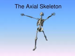
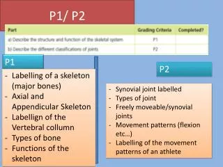
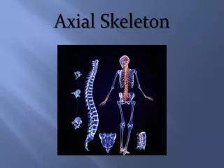
Chapter 7: The Axial Skeleton
Divisions of the Skeleton. Axial---bones that lie around the longitudinal axis of body.Appendicular

Chapter 7: The Axial Skeleton
E N D
Presentation Transcript
1. Chapter 7: The Axial Skeleton
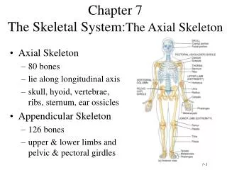
2. Divisions of the Skeleton Axial---bones that lie around the longitudinal axis of body.
Appendicular�bones of the upper and lower limbs plus bones forming the girdles.
3. Types of Bones Long bones
Have greater length than width
Has a shaft and variable number of extremities
Has mostly compact bone in the diaphysis and spongy bone in the epiphysis.
4. Types of Bones Short bones
Are equal in length and width (cube shape)
Is mostly spongy bone except at the surface which has compact bone.
Examples: Carpal bones of the wrist and tarsal bones of the ankle.
5. Types of Bones Flat bones
Are thin and composed of two parallel plates of compact bone enclosing a layer of spongy bone.
Provide protection and areas for muscle attachment.
Examples: Skull bones, sternum, ribs and scapulae
6. Types of Bones Irregular bones
Vary in size and shape.
Vary in the amount of compact and spongy bone.
Examples: Vertebrae, hip bones, facial bones, and calcaneus (heel bone).
7. Types of Bones Sesamoid bones
Develop in certain tendons to protect against friction, tensions and stress.
They are small in diameter (a few mm).
Change the direction of pull of a tendon.
Examples: patella
8. Types of Bones Sutural bones
Small bones located in sutures between cranial bones.
Number varies from person to person.
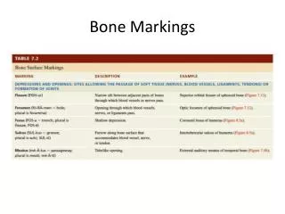
9. Bone Surface Markings Depressions and openings�used to form joints or allow the passage of soft tissues such as blood vessels and nerves.
Fissure�Narrow slit through which blood vessels and nerves can pass.
Fossa�shallow depression
Foramen�Opening through which blood vessels and nerves can pass.
Sulcus�furrow that accommodates a blood vessel, nerve or tendon.
Meatus�tubelike opening.
10. Fissures and foramens
11. Meatus
12. Sulcus and fossa
13. Bone Surface Markings Processes�projections or outgrowths that form joints
Condyle�large round protuberances
Facet�smooth flat articular surface
Head�rounded articular projection supported on the neck
14. Bone Surface Markings Processes�or serve as attachment points for connective tissue.
Spinous process
Crest
Tuberosity
15. Bone Surface Markings Processes�or serve as attachment points for connective tissue.
Epicondyle
Line
Trochanter
16. Bone Surface Markings Processes�or serve as attachment points for connective tissue.
Tubercle
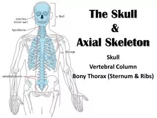
17. Skull bones Cranial bones�enclose and protect the brain
Facial bones�nasal (2), maxillae (2), zygomatic (2), mandible, lacrimal (2), palatine (2), inferior nasal conchae (2), and vomer.
18. Skull cavities Cranial cavity
Nasal cavity
Orbits
19. Cranial bones Frontal bones forms the forehead and the roof of the orbit.
Parietal bones form the sides.
Temporal bones form the inferior lateral part of the cranium
The mandibular fossa and articular tubercle articulate with the mandible to form the temperomandibular joint.
Houses the internal bones of the ear
20. Cranial bones
21. Cranial bones Occipital bone forms the posterior part and most of the base of the skull.
22. Cranial bones Sphenoid bone is the keystone the skull base, articulating with all the other cranial bones.
23. Cranial bones Ethmoid bone is a spongelike bone that forms:
Part of the anterior portion of cranial floor
Medial walls of the orbits
Superior portion of the nasal septum
Most of the superior walls of the nasal cavities.
24. Ethmoid bone
25. Facial bones Maxillae form the upper jaw bone.
Articulate with every bone of the face but the mandible.
Forms the bony roof of the mouth
Palatine bone form the posterior part of the hard palate
26. Facial bones Mandible makes up the lower jaw bone.
Only movable skull bone other than auditory ossicles.
The articulation of the condylar process with the mandibular fossa and articular tubercle of the temporal bone makes up the temperomandibular joint.
27. Facial bones Zygomatic bones�make up the cheekbones and part of lateral and floor of the orbits.
Lacrimal bones�smallest bones of the face that make up medial wall of orbits. Houses the lacrimal sacs.
Palatine bones�form posterior of hard palate and a small part of the orbital floors.
Inferior nasal conchae�make up part of the nasal cavity.
Vomer�a triangular bone that also makes up the floor of the nasal cavity.
28. Nasal cavity
29. Orbits The orbits house the eye and its structures.
Seven bones make up the orbit
30. Sutures Immovable joints in adults between skull bones. The joints can be movable in infants and children.
Coronal suture�unites frontal and parietal
Sagittal suture�unites both parietal
Lamboid suture�unites parietal and occipital
Squamous suture�unites parietal and temporal
31. Fontanels Mesenchyme filled spaces that have not ossified.
32. Hyoid bone A unique bone that does not articulate with any other bone. It is suspended from the hyoid processes of the temporal bone.
The hyoid bone and cartilages of the larynx are often fractured during strangulation.
33. Vertebral column Makes up to 2/5 of your height.
Is composed of a series of bones called vertebrae and of connective tissues (fibrocartilage).
It encloses and protects the spinal cord, which consists of nervous and connective tissues.
It supports the head, serves as a point of attachment for the ribs, pelvic girdle and back muscles.
34. The Vertebrae There are 33 vertebrae during early development and 26 as an adult.
7 cervical vertebrae
12 thoracic vertebrae
5 lumbar vertebrae
1 sacrum (5 fused sacral vertebrae)
1 coccyx (4 fused coccygeal vertebrae)
35. Intervertebral discs Found in between the vertebrae from the second cervical to the sacrum.
Consists of an outer ring of fibrocartilage called the annulus fibrosus and an inner soft, pulpy, highly elastic substance called nucleus pulposus.
36. Normal curves of the vertebral column Adult vertebral column has 4 curves
Convex cervical
Concave thoracic
Convex lumbar
Concave sacral
Curves increase strength, balance, absorb shock and protects against fractures.
37. Normal curves of the vertebral column Fetus has a single anteriorly concave curve
Primary curves-form during fetal development
Concave thoracic
Concave sacral
Secondary curves-form in the 10 months after birth.
Convex cervical
Convex lumbar
Curves are fully developed by age 10 but can be lost in old age.
38. Parts of a vertebra Body
Thick anterior portion
Has rough surface for attachment of intervertebral discs
Anterior and lateral surfaces contain nutrient foramina
39. Parts of a vertebra Vertebral arch--joins with the body to make up the vertebral foramen.
Pedicles
Laminae
The vertebral foramina make up the spinal cavity
40. Parts of a vertebra Vertebral notches�superior and inferior indentations in the pedicles
Stacking of vertebral notches form openings called intervertebral foramen, which allow passage of spinal nerves to a particular body segment.
41. Parts of a vertebra Processes that form points of attachment for muscles
Transverse
Spinous
42. Parts of a vertebra Processes that form joints with superior and inferior vertebrae
2 Superior articular processes
2 Inferior articular processes
Articulations are called intervertebral joints
43. Vertebral column regions: Cervical Have small bodies and large arches
Have 3 large foramina: one vertebral and two transverse.
The atlas (C1) is the first cervical vertebra inferior to the skull
The axis (C2)
44. Vertebral column regions: Cervical Atlas (C1)
Has no body but has anterior and posterior arches
Superior articular surface is concave and articulates with occipital condyles
Axis (C2)
The dens projects superiorly through vertebral foramen of axis
Vertebral prominens Atlas: Point out the pads on the atlas that let the head rest on the bones.
The atlanto-occipital joints permits you to nod yes
Axis � dens process � Body of the atlas is fused to the axis to form the dens (point out on the figure)
Forms a pivot for rotation of the skull and atlas allowing side to side motions
Children � have incomplete fusion between dens and atlas, so that impacts or severe shaking can cause severe damage to the spinal cord
In adults, a blow to the base of the skull can dislocate the atlanto-axis joint and drive the dens into the base of the brain � this can be fatal.
Vertebra prominens � last cervical vertebra. Interface between the cervical curve and thoracic curve
Has prominent spinous process that you can feel in your neck � the first of the spinous processes
Ligamentum nuchae � elastic ligament that stretches from the spinous process and attaches to the occipital bone of the skull � allows us to keep our head upright without expending any energy. Acts like bowstring. When you bend your head forward, it makes it easier to return your head to the upright position
Atlas: Point out the pads on the atlas that let the head rest on the bones.
The atlanto-occipital joints permits you to nod yes
Axis � dens process � Body of the atlas is fused to the axis to form the dens (point out on the figure)
Forms a pivot for rotation of the skull and atlas allowing side to side motions
Children � have incomplete fusion between dens and atlas, so that impacts or severe shaking can cause severe damage to the spinal cord
In adults, a blow to the base of the skull can dislocate the atlanto-axis joint and drive the dens into the base of the brain � this can be fatal.
Vertebra prominens � last cervical vertebra. Interface between the cervical curve and thoracic curve
Has prominent spinous process that you can feel in your neck � the first of the spinous processes
Ligamentum nuchae � elastic ligament that stretches from the spinous process and attaches to the occipital bone of the skull � allows us to keep our head upright without expending any energy. Acts like bowstring. When you bend your head forward, it makes it easier to return your head to the upright position
45. Vertebral column regions: Cervical Atlas: Point out the pads on the atlas that let the head rest on the bones.
The atlanto-occipital joints permits you to nod yes
Axis � dens process � Body of the atlas is fused to the axis to form the dens (point out on the figure)
Forms a pivot for rotation of the skull and atlas allowing side to side motions
Children � have incomplete fusion between dens and atlas, so that impacts or severe shaking can cause severe damage to the spinal cord
In adults, a blow to the base of the skull can dislocate the atlanto-axis joint and drive the dens into the base of the brain � this can be fatal.
Vertebra prominens � last cervical vertebra. Interface between the cervical curve and thoracic curve
Has prominent spinous process that you can feel in your neck � the first of the spinous processes
Ligamentum nuchae � elastic ligament that stretches from the spinous process and attaches to the occipital bone of the skull � allows us to keep our head upright without expending any energy. Acts like bowstring. When you bend your head forward, it makes it easier to return your head to the upright position
Atlas: Point out the pads on the atlas that let the head rest on the bones.
The atlanto-occipital joints permits you to nod yes
Axis � dens process � Body of the atlas is fused to the axis to form the dens (point out on the figure)
Forms a pivot for rotation of the skull and atlas allowing side to side motions
Children � have incomplete fusion between dens and atlas, so that impacts or severe shaking can cause severe damage to the spinal cord
In adults, a blow to the base of the skull can dislocate the atlanto-axis joint and drive the dens into the base of the brain � this can be fatal.
Vertebra prominens � last cervical vertebra. Interface between the cervical curve and thoracic curve
Has prominent spinous process that you can feel in your neck � the first of the spinous processes
Ligamentum nuchae � elastic ligament that stretches from the spinous process and attaches to the occipital bone of the skull � allows us to keep our head upright without expending any energy. Acts like bowstring. When you bend your head forward, it makes it easier to return your head to the upright position
46. Vertebral column regions: Cervical Atlas: Point out the pads on the atlas that let the head rest on the bones.
The atlanto-occipital joints permits you to nod yes
Axis � dens process � Body of the atlas is fused to the axis to form the dens (point out on the figure)
Forms a pivot for rotation of the skull and atlas allowing side to side motions
Children � have incomplete fusion between dens and atlas, so that impacts or severe shaking can cause severe damage to the spinal cord
In adults, a blow to the base of the skull can dislocate the atlanto-axis joint and drive the dens into the base of the brain � this can be fatal.
Vertebra prominens � last cervical vertebra. Interface between the cervical curve and thoracic curve
Has prominent spinous process that you can feel in your neck � the first of the spinous processes
Ligamentum nuchae � elastic ligament that stretches from the spinous process and attaches to the occipital bone of the skull � allows us to keep our head upright without expending any energy. Acts like bowstring. When you bend your head forward, it makes it easier to return your head to the upright position
Atlas: Point out the pads on the atlas that let the head rest on the bones.
The atlanto-occipital joints permits you to nod yes
Axis � dens process � Body of the atlas is fused to the axis to form the dens (point out on the figure)
Forms a pivot for rotation of the skull and atlas allowing side to side motions
Children � have incomplete fusion between dens and atlas, so that impacts or severe shaking can cause severe damage to the spinal cord
In adults, a blow to the base of the skull can dislocate the atlanto-axis joint and drive the dens into the base of the brain � this can be fatal.
Vertebra prominens � last cervical vertebra. Interface between the cervical curve and thoracic curve
Has prominent spinous process that you can feel in your neck � the first of the spinous processes
Ligamentum nuchae � elastic ligament that stretches from the spinous process and attaches to the occipital bone of the skull � allows us to keep our head upright without expending any energy. Acts like bowstring. When you bend your head forward, it makes it easier to return your head to the upright position
47. Vertebral column regions: Thoracic Are larger and longer than cervical vertebrae.
Spinous processes
T1 and T2
T11 and T12 Spinous processes
T1 and T2�long, laterally flattened and directed inferiorly
T11 and T12- shorter, broader and directed posteriorlySpinous processes
T1 and T2�long, laterally flattened and directed inferiorly
T11 and T12- shorter, broader and directed posteriorly
48. Vertebral column regions: Thoracic Articulate with tubercles of the ribs
Bodies have facets for articulating with heads of ribs.
Articulation with ribs is called vertebrocostal joint
Movement is limited by attachment of ribs to sternum
Spinous processes
T1 and T2�long, laterally flattened and directed inferiorly
T11 and T12- shorter, broader and directed posteriorlySpinous processes
T1 and T2�long, laterally flattened and directed inferiorly
T11 and T12- shorter, broader and directed posteriorly
49. Vertebral column regions: Thoracic Spinous processes
T1 and T2�long, laterally flattened and directed inferiorly
T11 and T12- shorter, broader and directed posteriorlySpinous processes
T1 and T2�long, laterally flattened and directed inferiorly
T11 and T12- shorter, broader and directed posteriorly
50. Vertebral column regions: Lumbar Are the largest strongest vertebrae
Have short thick processes
Superior articular processes are medial instead of superior
Inferior articular processes are lateral
51. Vertebral column regions: Lumbar Spinous processes project posterior
52. Vertebral column regions: Sacral Formed by the union of 5 sacral vertebrae
Serves as foundation for the pelvic girdle
Base�superior portion
Apex--narrow inferior portion Fusion starts between 16 and 18 and is completed by age 30.Fusion starts between 16 and 18 and is completed by age 30.
53. Vertebral column regions: Sacral Fusion starts between 16 and 18 and is completed by age 30.Fusion starts between 16 and 18 and is completed by age 30.
54. Vertebral column regions: Sacral Fusion starts between 16 and 18 and is completed by age 30.Fusion starts between 16 and 18 and is completed by age 30.
55. Vertebral column regions: Coccyx Formed by the union of 4 coccygeal vertebrae
Articulates with the apex of the sacrum
In females coccyx points inferiorly
In males coccyx points anteriorly Fusion starts later between age 20 and 30.Fusion starts later between age 20 and 30.
56. Bones of the thorax The thoracic cage is an enclosure made up of sternum, costal cartilages, ribs, and bodies of the thoracic vertebrae
Protects thoracic organs and provides support for pectoral girdle and upper limbs
57. Thoracic bones: Sternum Flat narrow bone
Manubrium is superior part
Body is middle part
Xiphoid process is the inferior smallest part-- does not ossify until age 40.
Fuses by age 25. Manubrium articulates with costal cartilages of 1st and 2nd ribs. Also articulates with the medial ends of the claviclesManubrium articulates with costal cartilages of 1st and 2nd ribs. Also articulates with the medial ends of the clavicles
58. Thoracic bones: Ribs 12 pairs, increase in length from 1 to 7
Each rib articulates with corresponding thoracic vertebrae
Ribs 1 to 7 (true ribs) articulate with sternum
Ribs 8-12 are false ribs
Ribs 11-12 are floating ribs.
59. Articulation of ribs with vertebrae Vertebrocostal joints�posterior portion connects to facet or demifacets of vertebra by its head and the rib tubercle connects to the transverse process.
60. Articulation of ribs with vertebrae
61. Rib injuries Breakage of ribs can happen at point of contact or weakest point (anterior to costal angle).
Dislocated ribs indicate displacement of costal cartilage from sternum.
Separated ribs are displace of a rib from its costal cartilage.
62. Temperomandibular joint syndrome (TMJ) Symptoms
Dull pain around the ear, headache
Tenderness of the jaw
Clicking or popping sound when the jaw is opened.
Limited opening of the mouth
Abnormal wearing of teeth.
Caused by improperly aligned teeth, grinding of teeth, arthritis or trauma to head or neck.