Download
1 / 72
830 likes | 1.7k Vues
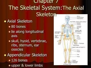
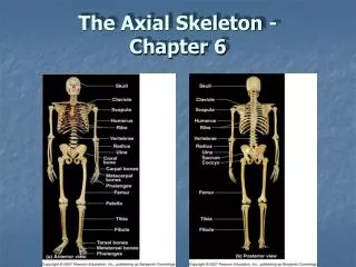
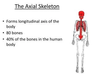
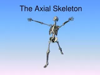
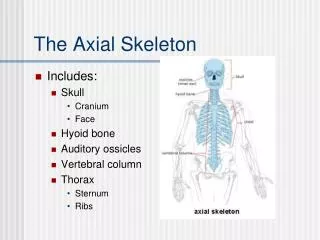
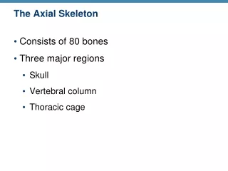
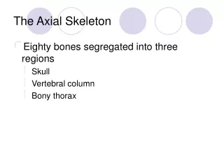
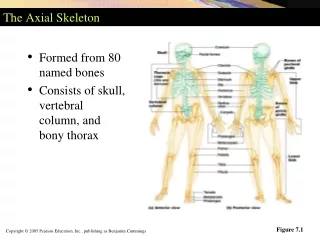
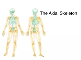
Chapter 7: The Axial Skeleton. Human Skeleton. Human Skeleton = 206 Bones Axial Skeleton : -longitudinal axis -80 bones Appendicular Skeleton : -limbs -126 bones. The Axial Skeleton. Figure 7–1a. Axial Skeleton. Appendicular Skeleton. Axial Skeleton Function.

E N D
Human Skeleton • Human Skeleton = 206 Bones • Axial Skeleton: -longitudinal axis -80 bones • Appendicular Skeleton: -limbs -126 bones
The Axial Skeleton Figure 7–1a
Axial Skeleton Function • Support and protect organs in dorsal and ventral body cavities • Provide surface area for muscle attachment: • Adjust position of head, neck, & trunk • Perform respiratory movements • Stabilize appendicular skeleton
Bones of the Axial Skeleton • The skull: 22 bones • 8 cranial bones: • form the braincase or cranium • 14 facial bones: • protect and support entrances to digestive and respiratory tracts • Skull bones interconnect at immovable joints called sutures • Dense fibrous CT
Cranial Bones • Enclose the cranial cavity • Which contains the brain: • and its fluids, blood vessels, nerves, and membranes
The Facial Bones • Superficial facial bones: • for muscle attachment • Maxillary, Lacrimal, Nasal, Zygomatic, and Mandible • Deep facial bones: • separate the oral and nasal cavities • form the nasal septum • Palatine bones, Inferior nasal conchae, and Vomer
The Maxillary Bones • The largest facial bones Figure 7–10a
Functions of the Maxillary Bones • Support upper teeth • Form inferior orbital rim • Form lateral margins of external nares • Form upper jaw and hard palate • Contain maxillary sinuses (largest sinuses)
The Palatine Bones Figure 7–10b,c
Functions of the Palatine Bones • Form the posterior portion of the hard palate • Contribute to the floors of the orbits
The Small Bones of the Face Figure 7–11
Functions of the Nasal Bones • Nasal Bones • Support the bridge of the nose • Connect to cartilages of the distal part of the nose (external nares) • Vomer • Forms the inferior portion of the bony nasal septum • Inferior Nasal Conchae • To create air turbulence in the nasal cavity • To increase the epithelial surface area • To warm and humidify inhaled air
The MandibleForms the lower jaw Figure 7–12a,b
The Hyoid Bone • Function: • Supports the larynx • Attaches muscles of the larynx, pharynx, and tongue Figure 7–12c
Marks of the Hyoid Bone • Greater horns (greater cornua): • support larynx • attach muscles of the tongue • Lesser horns (lesser cornua): • attach stylohyoid ligaments • support hyoid and larynx
Skull • Four major sutures: • Lambdoid: - separates occipital bone from parietal bones • Corona: - separates frontal bone from parietal bones • Sagittal: - separates parietal bones • Squamous: - (2) separates temporal bone from parietal bone
Sutures • The immovable joints of the skull Figure 7–3a, b
Sutures Figure 7–3c
Sutures Figure 7–3d, e
The Orbital Complex • Portions of 7 cranial and facial bones Figure 7–13
The Orbital Complex • Forms the eye sockets (orbits): • frontal bone (roof) • maxillary bone (floor) • maxillary, lacrimal and ethmoid bones (orbital rim and medial wall) • sphenoid and palatine bones
The Nasal Complex • Bones of the nasal cavities and paranasal sinuses Figure 7–14
The Nasal Complex: Sinuses • Sinuses: • air filled chambers inside flat bones • Function: • Reduce weight of bone • House mucus membranes that moisten and clean incoming air • Found in: • Sphenoid, ethmoid, frontal, palatine, and maxillary bones
The differences between the skulls of infants, children, and adults.
Skull Development • Intramembranous ossification from many centers of ossification • During development: • brain grows more rapidly than cranial bones • Growing skull bones are held together by bands of fibrous CT to provide flexibility • Expansion of brain, compression for birth • Large intersections of CT between the bones = fontalels (“soft spots”) • Persist until age 5 • Around age 5: • Brain stops growing in size, solid sutures form between cranial bones
The Infant Skull • Fusion is not complete at birth: • 2 frontal bones • 4 occipital bones • several sphenoid and temporal elements • Fontanels • Are areas of fibrous connective tissue (soft spots) • Cover unfused sutures in the infant skull • Allow the skull to flex during birth
The 4 Fontanels • Anterior fontanel: • frontal, sagittal, and coronal sutures • Occipital fontanel: • lambdoid and sagittal sutures • Sphenoidal fontanels: • squamous and coronal sutures • Mastoid fontanel: • squamous and lambdoid sutures
Skull Development Abnormalities • Craniostenosis: • Premature closure of frontanels, • Without surgery, the brain is crushed • Microcephaly: - Brain fails to enlarge - Cranium remains small
Craniostenosis Microcephaly
In which bone is the foramen magnum located? sphenoid occipital bone ethmoid parietal bone
Tomás suffers a blow to the skull that fractures the right superior lateral surface of his cranium. Which bone is fractured? frontal bone right temporal bone right parietal bone ethmoid
Which bone contains the depression called the sella turcica? What is located in this depression? sphenoid bone; pituitary gland ethmoid; olfactory epithelium temporal bone; inner ear lacrimal bone; tear apparatus
The vertebral regions, the curvatures of the vertebral column, and their functions.
The Vertebral Column: 26 Bones • The spine or vertebral column: • protects the spinal cord • supports the head and body • 7 cervical vertebrae (C1-C7) • 12 Thoracic vertebrae (T1-T12) • 5 Lumbar vertebrae (L1-L5) • 1 Sacrum (5 fused) • 1 Coccyx (3-5 fused)
Regions and Curves of the Vertebral Column • 26 bones: • 24 vertebrae, the sacrum, and coccyx • Vertebral column is not straight • 4 curves bring the weight of the body in line with the central axis Figure 7–16
The Vertebrae Figure 7–20a
Characteristics of the Sacrum and Coccyx • The sacrum: • is curved, more in males than in females • protects reproductive, urinary, and digestive organs • The coccyx: • attaches ligaments and a constricting muscle of the anus
4 Curvatures of the Vertebral Column • Cervical curve • Thoracic curve • Lumbar curve • Sacral curve
Primary Curves • Thoracic and sacral curves: • are called primary curves (present during fetal development) • or accommodation curves (accommodate internal organs)
Secondary Curves • Lumbar and cervical curves: • are called secondary curves (appear after birth in first year of life) • or compensation curves (shift body weight for upright posture) • Necessary for bipedalism • Cervical: holds head up • Lumbar: standing
Abnormalities in Curvature • Kyphosis: - exaggerated thoracic curvature • Lordosis: - exaggerated lumber curvature • Scoliosis: - abnormal lateral curvature
Construction of Column • Vertebral body: stacking • transfers weight along the spine • Intervertebral disc: • Spacing between bodies (not C1 and C2) • Annulus Fibrosus: Outside • Fibrocartilage • Nucleus pulposus: Inside • Gel (cushion) • Absorbs Shock • Loss of water from discs = shrinking height
Construction of Column • Elastic ligaments: • link bodies for alignment • Intervertebral foramen: • holes formed by spacing from discs, allow spinal nerves to exit column • Vertebral arch: • Bone attached to vertebral body, with body it forms vertebral foramen • Vertebral Foramen: • Hole for spinal cord • Vertebral Canal: • Bony canal for spinal cord • Formed by stacking of vertebral foramen