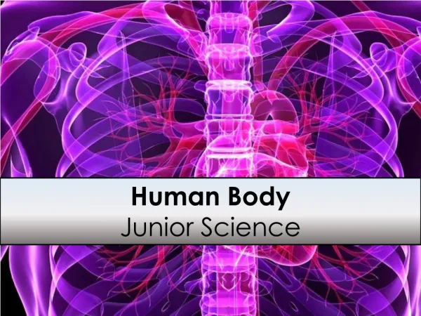
Human Body Junior Science
Human Body Junior Science. 1a. Is it Alive?. All living organisms are composed of cells. A cell is a small, membrane-bound compartment that contains all the chemicals and molecules that help support an organism's life. Biology is the study of living things.

Human Body Junior Science
E N D
Presentation Transcript
Human Body Junior Science
1a Is it Alive? All living organisms are composed of cells. A cell is a small, membrane-bound compartment that contains all the chemicals and molecules that help support an organism's life. Biology is the study of living things. A living object is an object that caries out life functions. A non-living object is an object that has not been alive. A dead object is an object that was once alive.
1a Is it alive? A twig once pulled off a tree is considered dead. A stick insect that looks a lot like a twig is alive, but how could you tell the difference? If you observe the twigs carefully, you should see the stick insect, move, eat food, and get rid of waste. The insect would be able to respond to the environment, for example it might move if something was brought up close to it, or a light was shone at the insect. If you waited long enough the insects in the tank might reproduce. The stick on the other hand would do none of these things.
1a All living things share the characteristics described in MRS GREN
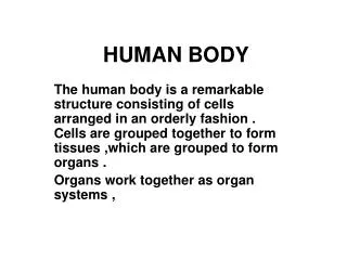
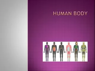
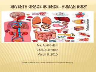
The structure and functions of the human (as an animal) systems: skeletal, digestive, circulatory, respiratory. 1b All vertebrates share the same basic body plan, with tissues and organs functioning in a similar manner. We focus on the human body, studying structure (anatomy) and function (physiology).
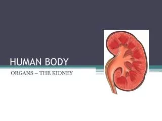
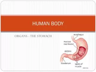
1b The body contains different organ systems Different tissues functioning together for a common purpose are called organs (eg, stomach, kidney, lung, heart). Organ systems are composed of individual organs working together to accomplish a coordinated activity. For example, the heart, veins and arteries all play a role in circulation.
1b The Human Organ Systems The Human Organ Systems that will be covered in this unit include the Cardiovascular system: including the Heart and associated blood vessels, the Skeletal system: Including the skeleton, muscles of the arms and legs and supporting connective tissue, and the Respiratory system: including breathing, gas exchange and respiration.
1c Organisation in the human body Organ Systems Body Organs The order of organisation in a body starts with organ systems (e.g digestive)that are made up of organs (e.g. stomach, liver) . Each organ is made up of one or more type of tissue (e.g. muscle) which in turn is made up of specialist cells. Each cell is comprised of smaller parts called organelles. Tissues Cells organelles
Extra for experts 1c The body is made up of cells Humans are made of complex systems of cells, which must be able to perform all of life’s processes and work in a coordinated fashion to maintain homeostasis (a stable internal environment). During a human’s early development, groups of cells specialize into three fundamental embryonic layers. These embryonic layers differentiate into a number of specialized cells and tissues. Tissues are groups of cells similar in structure and function .
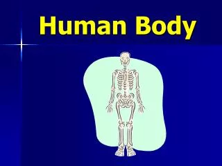
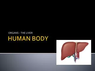
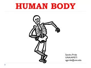
2a The Human skeletal system. The human skeletal system is an internal skeleton that serves as a framework for the body. This framework consists of many individual bones and cartilages. This also includes ligaments and the tendons which hold bones to each other and to muscles that contract and cause the bones to move. The adult human skeleton contains more than 200 bones. The skeleton also provides mechanical protection for many of the body's internal organs, reducing risk of injury to them, storage of minerals and production of blood cells.
2b The main bones in the Human skeletal system. The main bones of the human skeleton are divided into two groups. The axial skeleton is the central group of bones based around the spinal (vertebral) column. The appendicular skeleton consists of bones or groups of bones which “hang” off the axial skeleton.
2b The Human skeletal system. The main bones of the human skeleton are: The Skull Shoulder girdle - clavicle and scapula Arm- humerus, radius and ulna Hand - Carpals, Metacarpals and Phalanges Chest- Sternum and Ribs Spine- vertebrae, Sacrum (5 fused or stuck together bones) and Coccyx (the tiny bit at the bottom of the spine). Pelvic girdle - Ilium, Pubis and Ischium. Leg- Femur, Tibia and Fibula Ankle- Talus and calcaneus Foot - Tarsals, Metatarsals and Phalanges
2c Fractured Bones Extra for experts Bones are very strong and designed to handle a fair amount of impact and weight. Occasionally when the force is sudden and strong such as in a car accident or falling the bones will fracture. The type of fracture depends on how much damage is done, whether there is a crack (greenstick) or the bone is broken all the way through, as well as if it remains inside the body (closed)or breaks through the skin (open)
2c Ouch! How the body repairs Fractured Bones Extra for experts Most of the time the part of the body surrounding a broken bone is put into a cast to keep it still and allow the body to heal and repair the bone. So how does the body do this? >Immediately after the injury is flooded with natural painkillers called endorphins, which temporarily block out pain. >An injury then starts to swell because the body is sending extra oxygen and nutrients to the injury to begin the healing process. >A large hematoma, which is a collection of blood, surrounds the break in the bone. >Stem cells , which are responsible for making new cells, usually divide every one to two days. Now that there is an injury, they will divide every three minutes. >Within four weeks the hematoma will harden around the break, making the injured area extra strong. >Over the next several months, osteoclasts will "eat away" the hardened hematoma and the injury will be repaired. >Within a year of the injury, the bone will be almost as strong as it was before the break
Extra for experts 2d The structure of the skeleton and muscles of the human leg The two long bones in the leg provide a rigid frame that the muscles can pull against to provide movement. The thigh muscles attach across the knee joint etc. The muscles are attached to the bones by tendons. When the muscles contract they shorten and move the bones at the joints. All of the bones in the skeleton require muscles in order to move them. The muscles are attached by the nervous system to the brain which controls their movement – either voluntary or automatically when required
2d The structure of the skeleton and muscles of the human arm The muscles are attached to the bones by tendons. When the muscles contract they shorten and move the bones at the joints. All of the bones in the skeleton require muscles in order to move them. The muscles are attached by the nervous system to the brain which controls their movement – either voluntary or automatically when required.
The antagonistic muscles and bones act together to flex or extend the arm 2e Muscles can contract, but are not designed to actively lengthen, so they are arranged as opposing antagonistic pairs. As one muscle shortens, another is stretched and vice versa. The contacting muscles move the bones they are attached to.
2f A joint occurs where two bones meet . Movement of the skeleton occurs at the joints where two or more bones meet. There are different categories of joints. Freely movable, or synovial, joints allow a range of movement determined by the structure of the joint. Examples of movable joints are the ball and socket (shoulder), hinge (elbow), pivot (between the skull and neck) and ellipsoidaljoint (hand and arm). Ligamentsare inelastic connective tissues which hold bones together in a joint.
2f Other types of joint Extra for experts The two other groups of joints include the non-movable Fibrous joints and the moveable Cartilaginous joints. Fibrous joints are fixed in which no movement between the bones is possible. These joints do not allow movement because the bones are held firmly together by bundles of strong collagen fibres. Fibrous joints include the sutures of the skull bones and the peg and socket joints of the teeth in the jaw bone. Cartilaginous joints allow only slight movement. Often a pad of cartilage unites the two bones. An example of this is the joints between vertebral bones in the spine with cartilage discs between. Fibrous joints in the skull Cartilaginous joints in the vertebra
2g The synovial joint Cartilage is tough, rubbery surface that allows movement between bones, replaced by wear, lubricate the joint under pressure. The joint capsule is filled with a fluid to assist free movement of the joint. Ligaments hold the bones together and tendons hold the muscles to the bone.
2h Extra for experts Rheumatoid Arthritis – A case study Arthritis is inflammation of the joints (the points where bones meet) in one or more areas of the body. There are many different types of arthritis including Rheumatoid arthritis. Rheumatoid arthritis is a disease of the immune system, which normally protects us from infection by attacking viruses and bacteria. RA causes the immune system to mistakenly attack healthy cells such as the thin membrane that lines the joints. This causes fluid to build up in the joints, causing pain and inflammation. Over time this can wear away the cartilage and erode bone, causing a lack of function and mobility
2h Extra for experts Osteoarthritis– A case study Osteoarthritis is the most common form of arthritis and is most common in people over the age of 65. Osteoarthritis is a disease where there is a breakdown in the cartilage covering the ends of bones where they meet to form a joint and allow movement. As the cartilage wears away, the bones become exposed and rub against each other. The break down of cartilage also affects the shape and makeup of the joint so that it no longer functions smoothly.
The circulatory system 3a The structure of the circulatory system consists of blood, blood vessels and the heart. The function of the circulatory system is to be the body’s transportation system, moving oxygen, carbon dioxide, nutrients, wastes, hormones, vitamins, minerals and water throughout the body. It also aids in regulation of temperature.
3b Arteries carry blood away from the heart, veins carry blood towards the heart. Circulatory system A Deoxygenated blood enters the right atrium of the heart via the vena cava.B Deoxygenated blood is pumped from the right ventricle, via the pulmonary artery, to the lungs. C In the lungs, blood picks up oxygen and gets rid of carbon dioxide.D Oxygenated blood travels back to the heart in the pulmonary vein.E Oxygenated blood is pumped to vital organs and the whole body from the left ventricle, via the aorta.F Digested food is absorbed by blood through the walls of the small intestine.G Digested food is processed and stored in the liver.H Waste products (urea, water, salts etc.) are removed from the blood in the kidneys.I Blood supplies the rest of the body with food and oxygen. It carries away waste products.
The external and internal structure of the mammalian heart and its function. 3c The Human has a four chambered heart with full division between the ventricles. This means there is no mixing of the oxygenated and deoxygenated blood. It contains valves, and heart beat is controlled by the nervous system pacemaker.
The external and internal structure of the mammalian heart and its function. 3c The function of the Circulatory system must: >ensure delivery of blood to all tissues >adapt so that blood flow can be controlled and changed to individual tissues or the body as a whole >convert a pulsating blood flow in the arteries into a steady flow in the capillaries to allow optimum diffusion to and from the cells >return blood to the heart
Arteries carry blood away from the heart, veins carry blood towards the heart. 3d The vascular system is composed of arteries, arterioles, capillaries, venules and veins. The structure of the vessels in the different parts of the vascular system varies and the differences relate directly to the function of each type of vessel.
Capillaries link arteries with veins and are the sites of exchange with the tissues. 3d Arteries divide into smaller arterioles around targeted tissues. The arterioles divide once more into smaller capillaries which are only one cell thick. Nutrients and O2 diffuse across the membranes of the capillaries from the blood to the cell supplied – from high to low concentration. Waste products, such as CO2, will diffuse from the cell into the capillary near the venule end. The venules re-join into veins and waste product contained within the blood will be pumped back to the heart.
Arteries carry blood away from the heart, veins carry blood towards the heart. 3d There are three main types of blood vessels: >arteries, which carry blood away from the heart at relatively high pressure. >veins, which carry blood back to the heart at relatively low pressure. >capillaries which provide the link between the arterial and venous blood vessels Veinshave thinner, non-muscular walls to accommodate the lower blood pressure of the blood returning to the heart. Arterieshave thick muscular and elastic walls to accommodate the high blood pressure of the blood leaving the heart. Capillaries are only one cell thick.
Arteries carry blood away from the heart, veins carry blood towards the heart. 3d The Arteries and veins have different structures because they have different functions
Blood consists of fluid (serum) containing cells – white blood cells, red blood cells and platelets. 3e White blood cells (leucocytes) – some produce antibodies to tag foreign and harmful objects, others surround and eat tagged objects. White blood cells prevent infection becoming established. Make up the immunity system. Cells contain nucleus. Red Blood cells (erythrocytes) – contain haemoglobin, a pigment which binds to oxygen molecules and carries it through the circulatory system. They contain no nucleus. Platelets – they collect where there is a hole in the blood vessels and stop the unwanted flow of blood by clotting.
Red blood cells carry oxygen, attached to haemoglobin, around the body of a mammal. 3e Extra for experts Mammalian red blood cells (erythrocytes) dispose their nuclei and cell organelles when they are mature in order to provide more space for haemoglobin (oxygen carrying pigment). Because the cells have no nucleus or organelles, they cannot produce any RNA, and therefore they cannot divide or repair themselves. They are constantly replaced by new red blood cells. Red blood cells are biconcave disks and this shape optimises the cell for the exchange of oxygen in the lungs. The red blood cells are flexible so they can fit through tiny capillaries, where they release their oxygen to cells.
Extra for experts 3e Plasma transports glucose, carbon dioxide, urea and hormones. Plasma carries dissolved molecules required by the body to each cell and waste products like carbon dioxide and urea out of the body. Plasma also carries a large number of important proteins. Albumin, the main protein in blood, helps control the water content of tissues and blood. Plasma is usually yellow in colour due but can become milky when it transports fat absorbed from the intestines to other organs of the body.
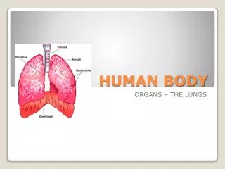
4a The respiratory system The respiratory system consists of organs that deliver oxygen to the circulatory system for transport to all body cells. The respiratory system also assists in removing the waste product of carbon dioxide from the body. Oxygen is a vital element for metabolism. The exchange of oxygen and carbon dioxide between body cells, blood and the air in the lungs is called respiration.
4a The structure of the Human Respiratory system The human respiratory system consists of the nose, pharynx, larynx, trachea, 2 bronchus, and lungs (composed of bronchioles and alveoli). The lungs are housed in an airtight pleural cavity framed by the rib cage and diaphragm.
4a The structure of the Human Respiratory system Air (comprised of about 21% oxygen) enters the nose, where tiny hairs filter out dust and particles. Tissues moisten and warm the air, making it more suitable for gas exchange in the lungs. Air passes from the pharynx to the larynx (containing vocal cords) and into the trachea.
4a The structure of the Human Respiratory system The trachea divides into the left and right bronchi which subdivide into smaller and smaller tubes called bronchioles. These airways are lined by mucous membranes and many cilia hairs which trap and remove particles from the lungs. Bronchioles open into the alveoli which are clustered like grapes.
4a Breathing, Gas Exchange and Respiration… What’s the difference? Breathing is the physical process by which air is forced into the lungs due to the ribcage and their associated muscles pulling the diaphragm downwards. The air is then forced out of the lungs by the same process that moves the diaphragm up. It involves Inhalation and Exhalation. Gas exchange is the process whereby gasses diffuse across a gaseous exchange surface. In humans, oxygen diffuses from the inhaled air into the blood and carbon dioxide diffuses from the blood into the exhaled air. The gaseous exchange surface in Humans is the Alveoli. Respiration is the metabolic process that occurs in the cell where organic molecules are broken down to release energy. In humans, this process takes place inside the mitochondria of cells. Aerobic respiration requires oxygen whereas anaerobic respiration takes place in absence of oxygen.
4b Breathing mechanism. Breathing (the movement of air in and out of the lungs) is produced by pressure differences created by changing the size of the pleural cavity. The diaphragm contracts and the rib cage is raised. The volume of the pleural cavity is increased, creating a partial vacuum between the lung cavity and the atmosphere, and air enters the lung.Air breathed out contains more carbon dioxide and less oxygen than air breathed in.
The structure of the alveoli and blood capillaries enable gaseous exchange to occur. 4c Only one cell thick, alveoli have direct contact with capillaries for gas exchange. The grapelike arrangement of alveoli creates an enormous surface area sufficient for exchanging enough oxygen and carbon dioxide for the entire body.
4c Gas exchange across the alveoli. Oxygen is transported to the cells of the body mainly by binding to haemoglobin which is found within red blood cells. Carbon dioxide is transported by diffusion into the plasma. Diffusion occurs because the oxygen is in higher concentration in the alveoli and moves to the lower concentration in the blood of the capillary. The CO2 in the blood diffuses into the alveoli and out of the body because it also moves from high to low concentration. Oxygen enters into alveoli from bronchioles The oxygen molecules must diffuse through both the lining of the alveolus and the lining of the blood capillary and is eventually picked up by red blood cells
Aerobic respiration involves transferring energy from glucose to a cell; oxygen is needed and carbon dioxide is produced. Extra for experts 4e Cellular respiration is a process whereby energy is released from the breakdown of molecules in food at the cellular level. Glucose enters the cell and oxygen diffuses in – which has previously entered the body via the respiratory system and transported to each cell through the circulatory system. Glucose is broken apart in a series of steps to release energy required for the body. Each reaction is assisted by enzymes. The products of these reactions are water and carbon dioxide – which diffuses back out of the cell and eventually out of the body via the circulation and respiratory systems.
Anaerobic respiration can occur in human muscles and that lactic acid is produced. Extra for experts 4f Sometimes our muscles require more oxygen than we can supply by normal breathing. Without oxygen present, anaerobic respiration takes place. The glucose is broken down and releases some energy. Carbon dioxide is produced along with lactic acid. The lactic acid is a waste product and must be removed from the body.
Extra for experts 4f Comparing aerobic and anaerobic respiration
The Big Picture Credit:http://thomasthinktank.pbworks.com/