Bronchus Suis: Anatomy Variants & Clinical Implications
Exploring the pig bronchus or "bronchus suis" anatomy, its common variants, and clinical implications such as impaired drainage, respiratory infections, and potential associations with other conditions like Down's syndrome. Understanding the normality of this anatomy in mammals like pigs and its significance in medical diagnosis and treatment.


Bronchus Suis: Anatomy Variants & Clinical Implications
E N D
Presentation Transcript
SPOTS Dr. Nehal Shah 1-12-07
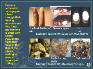
DIAGNOSIS "Bronchus Suis"or Pig bronchus with dissection of aorta D/D :- Diverticula of the right upper lobe bronchus (Diverticula lacks cartilaginous rings seen in tracheal bronchi.)
Right upper lobe bronchus arises directly from the lateral wall of the trachea prior to the bifurcation. FINDINGS
Of all bronchial anatomy variants, tracheal bronchus is one of the more common. It is almost always found on the right, supplying the apical segment of the right upper lobe. This anatomy is normal in other mammals such as pigs and therefore it is referred to as a "pig bronchus" or "bronchus suis". Although usually asymptomatic it may cause impaired drainage, respiratory infections, persistent coughing, stridor, bleeding, and bronchomalacia. It may be associated with tracheal hypoplasia or stenosis, lobar emphysema, pulmonary cysts, tracheo-esophageal-fistula, and has been seen in patients with Down’s syndrome. It can lead to right upper lobe atelectasis, especially in intubated patients.
Three variations of a tracheal bronchus have been described: The right tracheal bronchus may be a displaced bronchus with all three segments to the right upper lobe arising from it. In this variety there is no right upper lobe connection to the right main bronchus. The tracheal bronchus may consist of only a right upper lobe apical bronchus while anterior and posterior upper lobe bronchi arise from the right main bronchus. There is a supernumerary bronchus, the tracheal bronchus, leading to the right upper lobe in addition to normally structured (trifurcated) right main stem bronchus.
CPPD crystal diposition disease. Chondrocalsinosis
2 Elderly women with wrist and neck pain
11. K/C/O CA-ESOPHAGUS.
Findings The SVC is not present in its usual location to the right of the ascending aorta. There is an enhancing structure to the left of the aortic arch which is clearly a vessel. There is not normally any soft tissue structure (other than fat) in this location. This structure runs inferiorly to enter the coronary sinus, which on the third image is seen to course behind the left ventricle and drain into the right atrium.
Persistent left sided SVC, with absence of the right SVC. Discussion This variant occurs as a result of persistence of the left anterior cardinal vein. The right SVC often persists, but may be absent, as in this case. The incidence is approximately 0.5%. It typically drains via the coronary sinus.
Commonest of all vascular malformations of brain. CT scan: may be normal. may show popcorn calcification. absent / minimal enhancement MRI: complete Hemosiderin rim Multiple lesion are very common. Therefore, when one cavernous angioma is found, do GRE to detect other lesions. (GRE is more sensitive in lesion-detection, because of the blooming of the Hemosiderin rim, caused by magnetic susceptibility.)
Mediastinal lipomatosis It is a benign condition in which increased amounts of unencapsulated, histologically normal fat are seen in a variety of areas in the mediastinum. It has been associated with exogenous obesity, corticosteroid administration, and Cushing syndrome. On CT, the fat, which is homogeneous and similar in attenuation to subcutaneous fat (approximately -80 to -120 HU), most commonly is seen in the upper anterior mediastinum but also can be seen in the cardiophrenic angles and paraspinal region. Occasionally collections of fat can be seen in atrioventricular or interventricular grooves. If the fat is inhomogeneous, mediastinitis, neoplastic infiltration, or prior irradiation/surgery should be considered. However, small residual foci of thymic tissue should not be interpreted as being secondary to infiltration of the mediastinal fat
6. 30 year old female presented with chronic left knee pain. She had suffered minor trauma to the knee nine months previously.
MRI images (T1 sagittal, T2 sagittal, T1 coronal) show a well defined ossicle containing high signal marrow fat, lying in the posterior horn of the medial meniscus.
Diagnosis Meniscal ossicle Meniscal ossicles are a rare incidental finding. Usually found in the posterior horn of the medial meniscus, their etiology is uncertain. A congenital origin with the ossicle being formed from rests of primitive mesenchyme cells is preferred, but recent evidence supports a traumatic etiology.
The radiographs show a curvilinear fat containing lucency with an exostosis-like structure arising from the radius. The CT scan shows a lipoma with the exostosis in the centre of the lipoma. This appearance is characteristic of a parosteal lipoma.
Parosteal Lipoma It is a rare tumor, most common in the age group of 40-60 and forms 0.3% of all lipomas. It is most common in the thigh, but also seen in the upper extremity and other parts of the body. The exact site of origin of this lesion is uncertain. The appearance of a lipoma with a bony excrescence is classic for this tumor.
Ménétrier disease. Axial reformatted image shows large, lobulated folds and preserved gastric mucosa in the fundus.
Ménétrier disease is a rare chronic gastric disorder of unknown origin that predisposes for gastric cancer. It occurs most commonly in middle life, more often in men than in women. Grossly thickened lobulated folds of the gastric fundus and body are characteristic signs of Ménétrier disease, with relative antral sparing. The greatest degree of fold thickening occurs on or near the greater curvature. Focally enlarged folds can be mistaken for polypoid carcinomas.
10 Patient with a history of IV drug use and endocarditis.
Septic Pulmonary Emboli Multiple pulmonary masses, some of which are cavitating, are seen in both lungs and are compatible with septic puomonary emboli in this patient with a history of IV drug use and endocarditis.
11 This 58-year-old patient presented to the ER with breathlessness, productive cough, and fever.
Findings: A density is identified throughout much of the left lung. The left heart border is obscured. A crescent of air is seen around the aortic arch and lucency is seen at the left lung apex. Volume loss in the left hemithorax is appreciated. The right lung is clear, no infiltrate nor abnormal lucency is seen. The lateral view of the chest demonstrates increased density projecting anteriorly and superiorly in the chest projecting just superior to the heart and lying against the anterior chest wall.
12 h/o blunt trauma with insertion of ICD since 1 week.