SPOTS
Explore the case of Proximal Focal Femoral Deficiency (PFFD) affecting a baby's limb, with details on diagnosis, spectrum, and radiologic overview, along with other unique medical cases and conditions.


SPOTS
E N D
Presentation Transcript
SPOTS BHUMIKA SUTHAR SECOND YEAR RESIDENT SSGH,BARODA 11/04/08
1 Parents concerned about baby's unusually shaped limb
Findings: • The right femur is foreshortened with proximal dysplasia. • The proximal portion of the femur is subluxed laterally from the acetabulum. • The acetabulum is fairly well formed on the right.
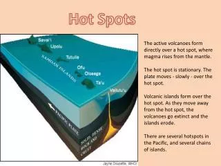
Proximal focal femoral deficiency
Discussion • Proximal focal femoral deficiency (PFFD) represents a spectrum of congenital hypoplasia or aplasia (deficiency) of the proximal femur. The abnormalities are usually centered roughly at the intertrochanteric femur. • This was associated with thalidomide use during the first trimester of pregnancy but most cases are of unknown etiology. • Radiologic Overview: • At the mild end of the spectrum, there is only hypoplasia of a short segment of intertrochanteric femur, normal femoral head and acetabulum. • A more severe case could have hypoplasia of the femoral head with acetabular dysplasia. • The most severe form involves absence of nearly the entire femur. Associated with absence of the ipsilateral fibula.
2. Hepatic artery angiography
POLY ARTERITIS NODOSA • Kidneys & liver commonly affected. • Affects medium & small sized arteries. • Selective angiography of renal artery or common hepatic artery show 2 to 3mm aneurysma with areas of arterial narrowing. • When these aneurysms thrombose, infarction of small areas of kidney produce Moth-eaten appearance on nephrogram.
Lace-like appearance of Sarcoidosis • In involve joints in 10%, causes transient migratory polyarticular arthrlgias without radiological findings. • A chronic granulomatous arthritis involving synovium. • Granuloma within or adjacent bone result in punched out cortical erosions or central lytic lesions with nonaggressive features within medullary cavity. • Fingers are the typical site. • Characteristic appearance caused by presence of multiple granulomatous lesions giving Lace-like appearance.
Congenital vertical talus • Congenital vertical talus or rocker-bottom foot is an uncommon rigid foot deformity which represents the most severe malformation on the spectrum of congenital flatfoot. • Congenital vertical talus, as well as oblique talus, is associated with several neuromuscular disorders which include arthrogryposis, neurofibromatosis, cerebral palsy, poliomyelitis, and spinal muscular atrophy. • the congenital vertical talus foot has a distinct plantar surface convexity, a rocker-bottom deformity & dorsal dislocation of navicular on talus.
Congenital Vertical Talus: A: An AP radiograph demonstrates an increased talocalcaneal angle due to the equinovalgus angulation of the os calcis.
DISH • Old people • Flowing osteophytes of the spine, involving 4 or more contiguous vertebrae. • Hyperostosis of ligamentous attachments. • In thoracic spine, the disease is less prominent on left side, due to aortic pulsations. • No SI joint involvement
CASE 7: 2 years old boy with convulsions & headache. NCCT CECT MRI IS NEXT..
D/D of posterior fossa lesions: * meduloblastoma : in children, with second peak in adulthood. * ependymoma: spreads along the ependymal tracks. * metastasis: most common post. Fossa lesion in adults. * pilocytic astrocytoma. * hemangioblastoma. • Hyperdensity on NCCT is a strong point in favour of meduloblastoma.
8. Pt came with h/o trauma
The radiographs show a curvilinear fat containing lucency with an exostosis-like structure arising from the radius. • The CT scan shows a lipoma with the exostosis in the centre of the lipoma. • This appearance is characteristic of a parosteal lipoma.
ParostealLipoma • It is a rare tumor, most common in the age group of 40-60 and forms 0.3% of all lipomas. • It is most common in the thigh, but also seen in the upper extremity and other parts of the body. The appearance of a lipoma with a bony excrescence is classic for this tumor.
CONNATAL CYST • These are cystic areas adjacent to the superolateral margins of the body and frontal horns of the lateral ventricles. • Connatal cysts are located at or just below the superolateral angles of the frontal horns or body of the lateral ventricles and are mainly anterior to the foramina of Monro. • These cysts have been reported to resolve at follow-up studies
SCLERODERMA OF BOWEL • “Hidebound" appearance on the inferior (mesenteric) border • CREST Syndrome • Calcinosis • Raynaud’s phenomenon • Esophageal dysfunction • Sclerodactyly • Telangiectasia
CASE-14 T- tube cholangiogram
Diagnosis: Caroli disease. • T-tube cholangiogram shows focal areas of saccular ectasia of the intrahepatic bile ducts. • The extrahepatic bile duct is normal.
Caroli Disease • Caroli disease is a congenital anomaly of the biliary tree that is characterized by saccular dilatation of the intrahepatic bile ducts and may be an autosomal recessive trait in some individuals. • The extrahepatic bile ducts are rarely involved, and there is no biliary obstruction. • The abnormality may affect only one hepatic segment or lobe, most commonly the left lobe. • Caroli disease is associated with autosomal recessive polycystic kidney disease and medullary sponge kidney. • Complications include cholangitis, hepatic abscess, and biliary stones. Recurrent inflammation leads to the development of cholangiocarcinoma in about 7% of cases. • Imaging shows scattered hepatic cysts communicating with the intrahepatic bile ducts.
Diagnosis- • Mucopolysaccharidoses(Hurler’s syndrome type-I)
Mucopolysaccharidoses(Hurler’s syndrome type-I) • Macrocephaly with mental retardation. • J- shaped sella. • Ovoid hook shaped vertebral body with thoracolumber gibbus ( inferior beaking). • Short wide phalanges with proximal pointing. • Iliac wings are flared with constricted base. • V-shaped deformity of metaphysis at wrist.
Lunate Dislocation • The normal PA view has three parallel arcs which aid in evaluation. • The first arc runs along the proximal articular margins of the proximal carpal row, and the second arc along the distal articular margins of the proximal carpal row. The third arc extends along the proximal articular margins of the distal carpal row. Continuity of these three arcs ensures carpal row integrity. In this particular case, arcs one and two are disrupted. This indicates a dislocation pattern. • The type of dislocation is easily detected on the lateral film. • Although the lateral film of the carpus presents as a confusing overlap of bones, it is fairly easy to pick out the coaxial relationship between the articular surfaces of the radius, lunate, capitate, and third metacarpal. • Although an exact linear relationship among these bones is uncommon, a coaxial relationship is essential. • In the current case, the lunate no longer articulates with the radius and the capitate no longer articulates with the lunate. The lunate is displaced volarly with 90 degrees rotation. The capitate remains aligned with the radius but sinks proximally. This is the classic configuration of a lunate dislocation.
PECTUS CARINATUM Lateral chest radiograph (a) and axial CT scan at level ofventricles (b) show characteristic anterior protrusion of the lower portion of sternum and the costal cartilages,with flattening of both sides of the chest.
ENOSTOSIS – typical brush border, increased radiotracer uptake.
Mafucci syndrome • Mafucci syndrome represents enchondromatosis with Soft tissue hemangiomas, usually in the hands and feet. • As with Ollier disease, there is typically a shortening of the long bones. These patients are at higher risk for sarcomatous transformation of both the vascular and cartilaginous portions of the disease.
20. Post Gd Coronal T1-W spin-echo MR image
Dural tail sign typically associated with Meningioma. • It was initially proposed that dural tails resulted from direct tumor invasion, but many later investigators were able to show little or no direct tumor involvement. • dural tails represented reactive changes to the dura mater, with perhaps minimal changes to meningothelial nodules that were adjacent to but not in contiguity with the tumor. • Meningiomas are known to be hyper vascular, which results in additional adjacent reactive changes such as hyperostosis and sinus blistering.