Download
1 / 55
580 likes | 833 Vues
Injuries to the Neck. Presley Regional Trauma Center Department of Surgery University of Tennessee Health Science Center Memphis, Tennessee. Assessment and Management. Introduction. Injuries to the neck can result from both blunt and penetrating trauma

E N D
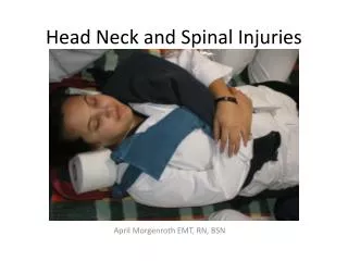
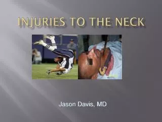
Injuries to the Neck Presley Regional Trauma Center Department of Surgery University of Tennessee Health Science Center Memphis, Tennessee
Introduction • Injuries to the neck can result from both blunt and penetrating trauma • Both mechanisms can cause devastating injuries • High associated morbidity and mortality • Penetrating injuries can be dramatic • Blunt injuries may present in a more subtle, delayed manner
Initial Management • Initial priority = ensure adequate airway • When in doubt, when all else fails … • Attention should be paid to signs of an injury • Patients with major airway injuries who are awake will position themselves to best protect their airway
Initial Management • Sit upright and lean forward • Keeps airway patent and allows for drainage of blood and secretions • DO NOT attempt to lie patient flat • May be better to go to OR for intubation with preparation for surgical airway • more controlled situation
Cricothyrotomy • Controversy over conversion • Traditional view (1921) – more likely to stricture • converted to trach on semi-elective basis within 24 to 48 hrs • 1976 – low complications with elective cric • Recent trauma studies show higher rate of complications for conversion
Initial Management • Management depends on depth of the wound, hemodynamic stability and the zone injured • Patients with penetrating injury and either hard signs of injury or hemodynamic instability require emergent operative exploration
Zones of the Neck • I – sternal notch to cricoid • trachea, great vessels, esophagus, thoracic duct, upper mediastinum and lung apices • II – cricoid to angle of mandible • carotid and vertebral arteries, jugular veins, esophagus, pharynx, trachea and larynx • III – angle of mandible to base of skull • distal extracranial carotid arteries, vertebral arteries and jugular veins
Initial Management • Those who do not require immediate exploration need a logical diagnostic approach to prevent a missed injury • Evaluation modalities are needed for each of the structures of the neck • Zones of the neck are useful as descriptors but not as a guide for management
Initial Management • Some are comfortable observing patients with a normal exam • Vascular studies are reserved for those who have soft signs of injury and hemodynamic stability • In contrast to extremities, missed injuries in the neck have more serious sequellae • justifying liberal use of angio and/or CTA
Management Principles • EARLY control of airway • No NGT • Angio for hemodynamically stable patients with zone I and III injuries • Hard signs and/or unstable = explore • Selective management vs mandatory neck exploration • with zone II – no difference in outcome
Operative vs Non-operative • The main factors influencing management decisions are: • hemodynamic stability • presence of hard signs • injury location
Hard Signs • Ongoing hemorrhage • Large or expanding hematoma • Bruit • Massive blood loss at scene • Hemiparesis or hemiplegia • Extensive subcutaneous emphysema • Stridor
Non-operative Evaluation • Reserved for patients with zone I or III injuries or zone II without hard signs • arteriography • pharyngoesophagoscopy • tracheobronchoscopy
Arteriography • Invaluable in excluding injury or planning operative approach for zone I or III injuries • Should visualize the innominate, carotid, subclavian and vertebrals for zone I - carotid and vertebrals for zones II and III • 4 vessel essential to exclude zone II injury with signs such as proximity, stable hematoma or h/o significant hemorrhage
Evaluation • Laryngoscopy • Flexible fiberoptic nasopharyngolaryngoscopy • Flexible bronchoscopy • Rigid or flexible esophagoscopy • Esophagram
Cervical Vascular Injuries • Neck trauma damages cervical vessels in 25% of cases • Penetrating trauma predominates • 30% have associated injuries in the neck and thorax • Blunt trauma accounts for < 10% of injuries • mortality rate = 10 – 30%
General Operative Approach • Active bleeding should be controlled with digital pressure until direct vascular control is achieved • Wounds should not be probed, cannulated or locally explored • these can dislodge clot and lead to uncontrolled hemorrhage or embolism
Operative Approach • Zone I - SCM incision + sternotomy • Zone II - SCM incision • Zone III - post-auricular extension with SCM incision + mandibular subluxation
SCM Incision • Provides exposure of the carotid sheath, pharynx and cervical esophagus • Can be lengthened to provide more extensive proximal or distal exposure • If bilateral exploration is necessary, separate incisions can be done
Management • Injuries to the CCA or ECA are governed by the extent of injury and overall status of pt • Simple injuries to the ECA should be repaired and complex injuries ligated • Complex injuries to CCA can be ligated if no neurologic deficits
Management • Injuries to the ICA are more problematic • Simple injuries with no interruption of flow should be repaired • Injuries to CCA or ICA with interrupted flow in the vessel, repair creates a theoretical disadvantage
Disadvantage • Interruption of flow may lead to focal brain ischemia and partial disruption of blood-brain barrier • Sudden restoration of blood flow may cause hemorrhage in the area of ischemia and worsen the extent of brain injury • Converted an ischemic infarct into a hemorrhagic infarct
To Ligate or Not to Ligate • Patient’s pre-op neurologic status • If there is no neuro deficit, it is presumed that there are no areas of brain ischemia • repair is safe • Focal neuro deficit is presumed to be related to ischemia • Clinical judgment • Milder neuro deficits may respond favorably to revascularization
Vertebral Artery Injury • Incidence • 1 – 7% with penetrating • < 1% with blunt • Mortality = 5% • Massive hemorrhage up to 15% • Neuro signs rarely found • Suspect with wounds posterior to SCM, facet joint dislocation or fracture through transverse foramen
Vertebral Artery Anatomy • V1 - subclavian origin to C6 TP foramen • V2 - interosseous portion C6 – C2 • V3 - C2 to foramen magnum • V4 - foramen magnum to basilar artery
Vertebral Artery Injury • Attempt angiographic diagnosis and therapy • Operate if hemodynamically unstable or for IR failure • Operative exposure is DIFFICULT
Exposure • Anterior SCM incision • Lateral margin of internal jugular vein is developed sharply and retracted medially • Proximal vertebral artery can be controlled after dissection deep to the supraclavicular fat pad
Tracheal Injury • Simple lacerations can be repaired in a single layer • Complex injuries can require tracheostomy • Extensive injuries can require delayed reconstruction • cartilage graft, fascial flaps
Tracheal Injuries • Avoid searching for recurrent laryngeal nerves • Separate tracheal and esophageal suture lines • Avoid tracheostomy through the repair • Flex the neck to avoid tension on the repair
Esophageal Injuries • Meticulous operative dissection must be employed to avoid missed injuries • Injuries should be debrided and repaired in two layers • Rotation of a muscle flap over the repair will decrease the incidence of fistula formation • Extensive injury may require cervical esophagostomy
Cerebrovascular Injuries • Blunt injuries to cervical arteries although rare can be difficult to diagnose and lead to devastating complications • True incidence is unknown • 1 to 2% of all blunt trauma patients, with higher rates in specific populations
Risk Factors • Signs/symptoms of injury • seat-belt mark, expanding hematoma, neurologic deficits • Mechanisms of injury • near-hanging • Specific head or neck injuries • Basilar skull fracture, cervical spine fracture, LeForte fracture
Carotid Trauma • No specific injury is pathognomonic • Direct compression between the angle of the mandible and the upper cervical vertebrae • Adbuction, extension and rotation of the neck causes stretch-traction injury • Initial injury is non-occlusive and serves as the focus for local thrombus formation and subsequent cerebral embolization
Presentation • Lucid interval • TIA, lateralizing sign • Cervical bruit • Neck contusion – 15% • Horner’s and/or anisocoria – 6%
Penetrating Injury Airway Control Unstable Zone II (Hard Signs) Immediate Exploration