NECK INJURIES
NECK INJURIES. Yolandé Smit. Perspective. 5 – 10% of all trauma cases 30% associated with injuries outside neck Leading cause of immediate death is exsanguination Esophageal injuries represents the most frequently missed injury and may be leading cause of delayed death
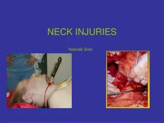

NECK INJURIES
E N D
Presentation Transcript
NECK INJURIES Yolandé Smit
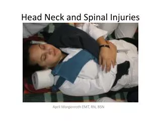
Perspective • 5 – 10% of all trauma cases • 30% associated with injuries outside neck • Leading cause of immediate death is exsanguination • Esophageal injuries represents the most frequently missed injury and may be leading cause of delayed death • Compound difficulties in evaluation & Mx is the complicated anatomy - dense concentration of vital structures in a small space • Ongoing debate : mandatory vs selective exploration
HISTORY • In 1552, Ambrose Pare ligated CCA. • WW1 – mortality 60% • WW11 - mandatory exploration, mortality 47% • Continuous advances in anaesthesia and peri-operative management – mortality 6% for early vs 35% for late exploration • Currently 2-6%
Mechanism of Injury • Penetrating • Blunt • Strangulation/ near-hanging
Penetrating Trauma (1) • 5-10% of all traumatic injuries • GSW • High velocity • Low velocity (50% lower incidence of significant lesions) • Stab wounds • Miscellaneous (shrapnel, impalement)
Location Arterial Venous Tracheolaryngeal Pharyngoesophegeal Spinal cord Neuroligical, other Thoracic duct Percentage % 12.8 11.3 10.1 9.6 3 3.4 0 Penetrating Trauma (2)Incidence Total 1275 patients
Blunt Trauma • Vascular injuries are rare but represent one of the most under diagnosed injuries • Aerodigestive injuries are rare in comparison to penetrating injuries but can cause acute AW compromise and delayed complications • Causes: • Motor vehicle collisions • ‘Clothesline’ injuries • Assault • Strangulation • Sports injuries
Strangulation • Near hanging • Complete/judicial • Incomplete • Typical • Atypical • Manual Strangulation • Ligature Strangulation • Postural Strangulation
Strangulation (2) Pathophysiology • High cervical fractures, complete cord transection, death • Venous congestion with stasis of cerebral blood flow leading to unconsciousness • Arterial occlusion with brain injury and death • Vagal reflexes contribute to fatal dysrythmias • Pulmonary sequelae Compression of AW doesn’t play as significant role in incomplete hanging as does vascular occl
Anatomy Three components • Structural • Visceral collumn • Respiratory layer (trachea & larynx) • Alimentary layer (pharynx & esophagus) • Endocrine layer (thyroid & parathyroid) • Paired neurovascular bundle
Anatomy Two fascial layers • Superficial fascia • Beneath skin • Contains platysma • Deep fascia • Investing layer • Visceral layer • Prevertebral
AnatomyPrevertebral Fascia • Covers paraspinal structural components • Two layers/laminae: alar & prevertebral • Fans out to cover roots of brachial plexus and subclavian a • Axillary sheath
AnatomyVisceral Fascia • Forms visceral compartment • Pretracheal anteriorly • Buccopharyngeal and retroesophageal posteriorly • Portion enclosing the strap muscles a.k.a middle cervical fascia • Space between buccopharyngeal and prevertebral fascia
AnatomyInvesting fascia • Envelopes Trapezius & SCM • Forms complete sheath of neck • Base of skull to sternum • Suprasternal space
AnatomyCarotid Sheath • Loose aggregation of connective tissue • Visceral compartment medial • SCM anterolateral • Prevertebral fascia posteriorly
AnatomyFascial Layers: Importance • Superficial layer contains platysma: important surgical landmark • Tight fascial compartments may limit external hemorrhage from vascular injuries BUT easily compromises the airway • Danger space: mediastinits
AnatomyThree Zones • ZONE 1- thoracic outlet • Cricoid cartilage to sternal notch • ZONE 2- Central • Cricoid to angle of mandible • ZONE 3 - Skull base • Angle of mandible to base of skull
AnatomyZones • Zones apply to neck anterior to anterior border of trapezius • Wounds to posterior triangle rarely associated with vascular, airway or digestive injury, except vertebral artery • Wounds through SCM or anterior triangle have high likelihood of injury • Decision making for diagnostic tests & surgical approach
AnatomyBrachiocehalic A • 4-5 cm in length • Ascend obliquely to right of sternoclavicular joint • Divides into right common carotid and subclavian aa
AnatomySuclavian A • Passes behind SCM, Jugular vein and Vagus nerve • Three branches • Vertebral • Internal mammary • Thyrocervical trunk • Courses post to anterior scalene than crosses first rib to become axillary artery
AnatomyCarotid A • Common carotid has no branches • Bifurcates to external and internal carotids at superior border of thyroid cartilage • Internal – no cervical branches • External – 8 branches
AnatomyVertebral A • First branch of subclavian • Extra-osseous until entering the transverse process of C6 (V1) • Intra-osseous from C6 – C2 (V2) • Distal extracranial top of C2 to base of skull (V3)
Initial ManagementAirway (1) Indications for immediate intubation • Apnoeic or near apnoeic patient • Comatose patient • Significant respiratory compromise (stridor, dysphonic with air hunger) • Rapidly expanding neck hematoma • Massive subcutaneous emphysema causing airway compression or distortion of trachea or larynx • Massive bleeding into airway
Initial ManagementAirway (2) ‘Wait-and-see’ • If no immediate indication • Fastidious observation of clinical status • Equipment readily available • Prompt intubation if any evidence of expanding hematoma or enlarging neck sc emphysema
Initial ManagementAirway (3) • Avoid BMV if possible • Oral intubation preferred • Without sedative in unconscious/ flaccid/near apnoeic patient • With sedation in uncooperative/agitated pt • RSI technique-of-choice • Difficult intubation anticipated: • ‘Brutane’ technique • Ketamine 1-2mg/kg IV
Initial ManagementAirway (4) • Surgical airway last resort • Cricothyrotomy preferred to tracheostomy: • Time • Position of neck • Bleeding • If laryngeal injury suspected • Consider awake local tracheostomy • Cricothyroidotomy for emergencies • Fibreoptic laryngoscope only if prepared to emergently obtain surgical airway • Needle cricothyroidotomy + jet insufflation may be acceptable to temporize • Retrograde intubation and blind nasal intubation is contra-indicated
Initial ManagementAirway (5) Pediatric considerations • Ideally with RSI • Fewer AW salvage techniques available: • Cricothyrotomy contra-indicated under 10 • Emergency tracheostomy difficult • Degree of AW obstruction after blunt trauma to larynx inversely related to degree of calcification, putting children at highest risk
Initial ManagementControl bleeding • Local pressure • Proximal and distal control • Balloon tamponade with Foley’s catheter • Two catheters in supraclavicular zone1 • Immediate ED thoracotomy with aspiration of air from RV if cardiac arrest immediately before or after arrival in ED
Initial ManagementPitfalls • Do notprobe wounds • Platysma penetration mandates evaluation in controlled fashion • Controversy about NGT • Tube may migrate from esophagus • Valsalva by patient may dislodge clot • Bloodstained
Initial ManagementImmediate Surgical Exploration INDICATIONS • Severe active bleeding • Shock not responding to fluids • Rapidly expanding hematoma • Absent radial pulse • Evolving stroke
Initial ManagementComplete Primary Survey • Thorough examination • Vascular: Hard signs of vascular injury • Airway: Stridor hoarseness, dyspnoea, sc emhysema
Initial ManagementSecondary survey • Digestive tract • Dysphagia • Odynophagia • Hematemesis • Subcutaneous emphysema • Neurological: focal signs
Further Management • If no indication for immediate surgical exploration, subsequent intervention based on : • clinical exam • Hard signs • Soft signs • No symptoms or signs • diagnostic testing
Hard Signs • Pulsatile bleeding • Expanding hematoma • Not responding to resus • Absent distal pulses • Distal signs • Cold, pale limb • Stroke • Bruit • Airway obstruction
Soft signs • Venous bleeding • Stable hematoma • Responding to resus • Diminished peripheral pulses • Proximity to major artery • Peripheral nerve deficit (brachial plexus) • Hemoptysis/hematemesis • Dysphonia/dysphagia
Diagnostic Strategies • Stable patient with hard signs: exploration (diagnostic strategies to plan) • Stable patient with soft signs/ asymptomatic: mandatory vs selective exploration (diagnostic strategies to aid in diagnosis) • Is physical examination reliable? Consider: • Resources • Potential for injury • Ability to serially exam • Consequences of missed injury
Diagnostic strategies • X-Rays • Chest: PT/HT, widened mediastinum, mediastinal air, elevated hemidiaphragm, FB • Neck: prevertebral air or swelling, sc air, FB • Gastrograffin swallow • All penetrating neck injuries due to high incidence of occult injuries • Endoscopy • If swallow (-), enhances sensitivity for penetrating esophageal injury
Diagnostic Strategies • Arteriogram • Diagnostic • To plan surgery • Therapeutic • Laryngoscopy/ bronchoscopy • Hemoptysis/ hoarseness/ sc emphysema/ laryngeal tenderness or deformity • CT neck • Useful for evaluating laryngeal injuries • May play a role in the future as screening modality in diagnostic evaluation of AW & vascular injuries in asymptomatic pt
Diagnostic strategies Airway/resus/assessment Stable Unstable Active hemorrhage Expanding hematoma Evolving stroke Zone 1 Zone 2 Zone 3 Swallow Endoscopy Arteriogram Arteriogram Swallow Endoscopy Explore + - - + Observe Explore Embolize Observe