Download
1 / 61
610 likes | 1.26k Vues
Penetrating and Blunt Neck Injuries “Deadly Missed Injuries”. Bradley J. Phillips, MD Burn-Trauma-ICU Adults & Pediatrics. Types of Injury - Penetrating. 40% do not involve important structure GSW direct and delayed type of injury Structures major vein 15-25% major artery 10-15%

E N D
Penetrating and Blunt Neck Injuries“Deadly Missed Injuries” Bradley J. Phillips, MD Burn-Trauma-ICU Adults & Pediatrics
Types of Injury - Penetrating • 40% do not involve important structure • GSW direct and delayed type of injury • Structures • major vein 15-25% • major artery 10-15% • pharynx or esophagus 5-15% • larynx or trachea 4-12% • major nerves 3-8%
Type of Injury - Blunt • Cervical spine • Vascular injuries • internal carotid artery • vertebral carotid artery • Aerodigestive • esophageal (rare) • larynx
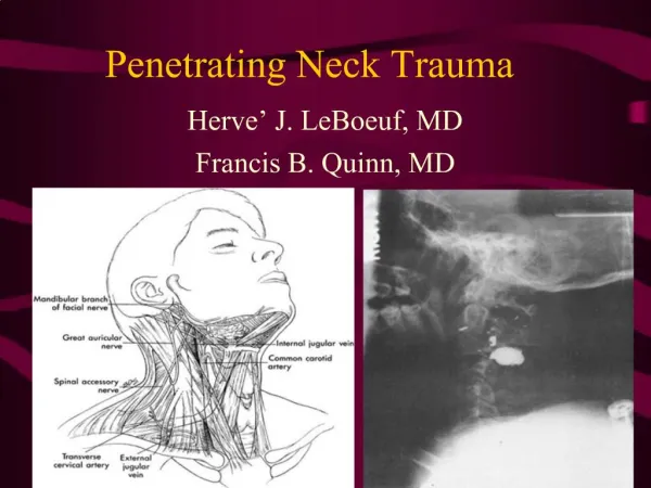
Deadly Missed Neck Injuries • Carotid Artery Injury • Esophageal Perforation • Laryngotracheal Injury
Diagnosis • Significant injuries often asymptomatic • 25% positive symptoms and 25% positive signs • PE is often deceptively negative for severe injury • Symptoms variable and delayed • internal carotid artery > 2 weeks • esophageal • Weigelt (A J Surg 1987) 3/10 no signs or symptoms • laryngeal • more likely to have presenting symptoms/signs • voice change, SOB, hemoptysis
Keys to Diagnosis • Little need for labs • High index of suspicion • Sense of urgency • Operation vs radiology
Case #1 • 21 yom with GSW to right neck without exit site • c/o pain in throat/right neck • VS : HR 110, BP 130/70, RR 27 sats 98% (40%) • PE: • mild swelling right neck, non-pulsetile • ??
Vascular Injuries - Physical Exam • Penetrating • Fogelman et al (Am J Surg,1956) • 43% hemodynamically stable • 70% no sign of bleeding • Carducci et al(Ann Emerg Med, 1985) • 1/3 no signs/symptom • Apffelstaedt et al (World J Surg, 1994) • Prospective study, 335 patients • SW penetrating platysma • clinical signs absent 30% of positive neck explorations
Physical Exam - Penetrating • Reliable for significant vascular injuries • Demetriades et al (Br J Surg, 1993) • prospecitive 335 patients, detailed written protocol • 7/335 required angiography • 269/335 nonoperative managed • 2 required subsequent operations for vascular injury • no complications • Demetriades et al (World J Surg, 1996) • prospective 223 patients, strict written protocol(Doppler) • 160/223 no clinical signs underwent angio • no vascular injury requiring treatment
Overview Management Penetrating Neck • Zone I • routine angiography • ? Esophageal evaluation (EGD, swallow) • Airway evaluation (bronchoscopy) • Zone II • selective management vs operative • neither approach superior (Asensio et al, Surg Clin N Amer, 1991) • Zone III • routine angiography
Angiography • Recommended in Zone I and III • difficult to assess clinically • difficulty surgical exploration • Policy reduces nontherapeutic intervention • Costs (Demetriades et al, Br J Surg, 1993) • Zone I only 5% required operation • Zone III only 13% required operation
Management Penetrating Zone II • Mandatory exploration • Advantages • decreased injuries • up to 25% unexpected injuries found • low morbidity/mortality • Disadvantages • report up 67% negative exploration • Recommendations • Zone II injuries with/without instability • GSW that cross midline
Transcervical GSW • More likely to involve vital structures • 73% vs 31% (GSW not cross midline) • Hirshberg et al, Am J Surg 1994 • retrospective 41 patients • 30(83%) positive for cervical injury • recommends mandatory exploration • Demetriades et al, J of Trauma, 1997 • prospective, 33 patients • 73% injury to vital organ, only 21% therapeutic operation
Stab vs Gunshot Wounds • Anecdotal suggestion • explore GSW, non-operative SW • not supported in literature • Prospective study (Demetriades et al, Br J Surg, 1993) • 97 GSW, 89 SW • GSW higher incidence of clinical signs than knives (35% vs 19%) • GSW more likely injuries • therapeutic operation: GSW 16.5%, SW 10.1%
Zone II - “Selective Conservatism” • If hemodynamically stable • angiography, contrast study, endoscopy , +/- laryngoscopy • Exploration if positive study • Negative neck exploration 20% • Missed injuries negligible (Jurkovich et al, Trauma, 1985) • Disadvantages • cost and time • iatrogenic (CVA, esophageal perfs)
Treatment- Specific Injuries • Carotid injuries • 22% of penetrating cervical vascular injuries • mortality 10-20% (in-hospital) • Repair vs ligation • repair if possible in absence of neurologic deficits • prefer saphenous vein, but prosthetics ok • if internal carotid injuries, transposition of external carotid • ligation in neurologically intact for high internal carotid injury if very difficult or impossible
Treatment- Specific Injuries • Carotid injury • Presence of neurologic deficits • controversial • ? Concern of postvascularization hemorrhagic infarct • increased risk if evidence of sever anemic infarct or edema • recommend repair • if deficits are short of coma • no evidence of anemic infarct • patent distal carotid
Treatment- Specific Injuries • Carotid artery occlusion with symptoms • may result in late local or neurologic complications • may develop pseudoaneurysm or rupture • recommend repair if • technically feasible • not at base of skull
Treatment - Specific Injuries • Minor carotid injuries (intimal flaps) • natural history not known • controversial: observation vs aggressive approach • ? role of duplex for decision making • role of anti-platelet unproven, but used
Management - Specific Injuries • Vertebral artery • increased frequency secondary liberal angio • 10% of major vascular injuries • 67% have association with major cervical injury mainly spine • isolate injury asymptomatic in 1/3 patients • thrombosis rarely lead to neurologic sequelae • angiographic embolization standard of care if bleeding
Complications • Nonoperative Management • delayed bleeding • CVA (dissection, emboli) • pseudoaneurysm • sepsis (missed esophageal leak) • Operative Management • injury to nerves (vagus, hypoglossal, recurrent) • blood loss • missed injury (particularly esophageal)
Summary Treatment - Vascular Injury • Surgical exploration unstable and stable Zone II (board answer) • Angiography Zone I and III • ? Nonoperative management stable Zone II • depends on expertise and facilities • Other interventions • embolization high carotid or vertebral artery • endovascular stent (pseudoaneurysms) • anticoagulation blunt carotid/vertebral artery
Case #2 • 56 yom s/p MVC driver vs pole • Found unconscious at scene, intubated • VS: HR 90, BP 110/80, sat 100% • PE: • abrasions to left shoulder/mid chest/LUQ • GCS 7, pupil equal/reactive • ??
Carotid Artery Dissection Internal Carotid Occlusion
Blunt Carotid Injury • Low incidence (0.08-0.25%) • Male 76%, Mean age 35 +/- 2 yrs • Most commonly intimal disruption • ? asymptomatic • Louisville U. (1998) 24 BCI all symptomatic • Colorado U. (1998) 12/56 asymptomatic • Often delayed diagnosis (Krajewski, Ann Surg 1980) • 58% > 10hrs • 36% > 24 hrs
Eiology MVC 41% (seat belt not a factor) Fall/ped struck 14% MCC 11% other 22% ski bike assault near hanging horseback Associated injuries CHI 65% facial fx 60% thoracic 51% basilar skull fx 32% extremity fx 32% abdominal 30% pelvic fx 16% cervical fx 5% none 16% Blunt Carotid Injury Biffl et al, Ann Surg, 1998
Diagnosis - Vascular Injury • Careful PE • hematomas, bruit, thrill • Horner’s syndrome • limb paresis or paralysis • deep coma • Delayed up to several days • PITFALL: Failure to consider blunt carotid injury with negative CT and CNS changes delayed
Blunt Carotid Injury • Screening asymptomatic (Biffl et al, 1998) • severe neck hyperflexion, flexion, or rotation • significant soft-tissue injury anterior neck • cervical spine fracture • displaced midface fx or mandibular fx associated with a major injury mechanism • basilar skull fx involving sphenoid/mastoid/petrous/foramen lacerum
Blunt Carotid Injury • Biffl et al, 1998 (continued) • before screening 12/12429 (0.1%) • after screening 25/2902 (0.86%) • only 28% had lateralizing signs/symptoms • 25% had concomitant head injury/depressed MS • symptoms and timing • > 24 hrs - 28% • > 1 week - 8.3 %
Blunt Carotid Injury • Biffl et al, 1998 (continued) • Outcome by symptoms at diagnosis • Dead Major Minor Normal • Asymptomatic 1 1 0 11 • Symptomatic 7 6 5 6
Blunt Carotid Injury • Biffl et al, 1998 • Treatment • Operative =1/37 • Anticoagulation = 24/37 • endovascular stent 10/24 • No intervention = 11/37 • Outcome • Dead Major Minor Normal • Anticoagulation 1 6 4 13 • No Anticoagulation 3 1 1 4
Blunt Carotid Injury • Biffl et al, 1998 • Complications • angiography (2) - groin hematoma(1), CVA (1) • hemorrhagic • 54% rebleeding ( transfusions and/or cessation) • Summary • Anticoagulation improves outcome • confirmed Fabian et al, Ann Surg, 1996 • Aggressive screening ( ? Diagnostic test) • Optimal intervention ? • Stenting pseudoaneurysm
Blunt Carotid Injury Contraindicaton to Heparin No Yes Heparin Observe Angiography 7-10 d Pseudoaneurysm Yes No Coumadin 3 mos Heparin/Stent or OR
Vascular Injury - Radiologic Test • C- spine films • r/o fractures (spine/larynx) • ? subcutaneous air • anterior cervical soft tissue swelling • tracheal deviation • ? CXR/ skull xray (where’s the bullet ?)
BCI and Anticoagulation Fabien et al, Surg, 1996
Vascular Injury - Radiologic Test • Duplex • can be used for carotid injuries Zone II only • as useful as angio in stable patients with Zone II injury (Thal, Trauma, 1991) • operator dependent • CT Angiogram • limited studies • ? comparable to angiogram (missed blunt injuries) • advantage: no risk of CVA
Vascular Injury - Radiologic Test • Angiography • gold standard (4 vessel runoff) • Indications • proximity to carotid with or without hematoma • shotgun blasts with ? multiple artery segments injuries • precise localization for planning proximal or high carotid injury • blunt trauma with extensive soft-tissue injury • blunt trauma with neurologic loss unexplained by CT
Case #3 • 29 yof restrained driver, head-on MVC • reported striking face/neck on steering wheel c/o neck/throat pain • airway patent without voice change • PE: • anterior neck crepitus • no stridor • ??
Diagnosis - Esophageal • Blunt esophageal injury rare • High index of suspicion in blunt trauma • Penetrating trauma - evaluation part of a complete work-up • If missed, high morbidity/mortality
Esophageal Injury - Diagnostic Test • Contrast swallow • Extravasation is diagnostic • Negative study is not reliable (particular in neck with gastrograffin) • 50% of leaks missed with gastrograffin • 25% of leaks missed with barium
Esophageal Injury - Diagnostic Tests • Controversy of initial contrast to use • gastrograffin • pneumonitis if aspirated • barium • increased inflammation/infection in the mediastium • Rec: If gastrograffin study is negative, repeat swallow this barium. Avoid gastrograffin in patients without gag/cough
Esophageal Injury - Diagnostic Test • Endoscopy • Generally recommended when contrast swallow is negative, but suspicion is high • Perforations often readily seen, however • 50% missed (Weigelt et al Am J Surg 1987) • missed in pharynx and cervical esophagus • missed in patients on ventilator (poor expansion of esophagus) • Combination of swallow/esophagoscopy reduces missed injuries to < 5%