Penetrating and Blunt Neck Trauma Tintinalli Chapter 258
Penetrating and Blunt Neck Trauma Tintinalli Chapter 258. Anatomy. Complex network of neurovasuclar & muscular structures supported by various fascial planes Two methods used to describe the neck: Zones & Triangles


Penetrating and Blunt Neck Trauma Tintinalli Chapter 258
E N D
Presentation Transcript
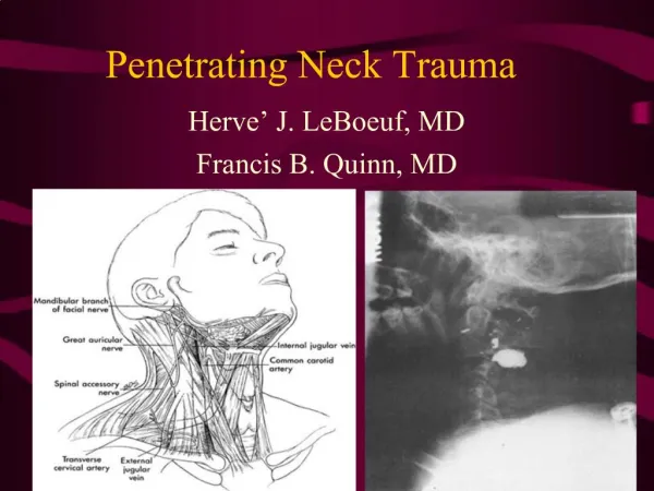
Anatomy • Complex network of neurovasuclar & muscular structures supported by various fascial planes • Two methods used to describe the neck: Zones & Triangles • Anterior Triangle: anteriorly by the midline; posteriorly by the SCM; superiorly by the mandible • Posterior Triangle: anteriorly by the SCM; inferiorly by the clavicle; posteriorly by the anterior border of the trapezius muscle
Anatomy • Anterior Triangle Structures: carotid artery, internal jugular vein, vagus nerve, thyroid gland, larynx, trachea, and esophagus • Posterior Triangle Structures: few vital structures, except at its base, where the subclavian artery and brachial plexus are located
Anatomy • Zones • One: superiorly from the sternal notch & clavicles to the cricoid cartilage (injury affects both neck & mediastinal structures) • Two: cricoid cartilage to the angle of the mandible • Three: angle of the mandible to the base of the skull
Anatomy • Zone I: includes the vertebral and proximal carotid arteries, major thoracic vessels, superior mediastinum, lungs, esophagus, trachea, thoracic duct, and spinal cord • Zone II: involve the carotid and vertebral arteries, jugular veins, esophagus, trachea, larynx, and spinal cord • Zone III: includes the distal carotid and vertebral arteries, pharynx, and spinal cord
Anatomy • Fascial Planes • Platysma: thin muscle covers the entire anterior triangle and the anteroinferior aspect of the posterior triangle; serves as an important planar landmark when evaluating penetrating neck injuries • Deep Cervical Fascia: invest deep structures; important due to the pretracheal deep fascia’s communication to the anterior mediastinum (neck trauma can lead to mediastinitis)
Initial Management • ABC’s • Always be ready for a cricothyroidotomy • Maintain C Spine alignment until cleared via Nexus (if you believe in it) or imaging or BOTH • Direct pressure for bleeding • Disability: try to assess quickly if intubation
Hard (trauma more likely) Hypotension in the ED Active Arterial Bleeding Diminished Carotid Pulse Expanding Hematoma Bruit Lateralizing Signs Hemothroax > 1000 mL Air Bubbling Wound Hemoptysis Hematemesis Soft Hypotension in the field HX of Arterial bleeding Tracheal deviation Large hematoma Stridor Hoarseness Vocal cord paralysis Sub Q Air 7th cranial nerve injury Unexplained bradycardia Signs and Symptoms
Diagnostic Strategies • Prospective study of 393 patients with penetrating neck injuries found 30% of patients with no physical findings had positive findings on surgical exploration. • Another study of 223 patients found physical examination was reliable to determine which patients needed vascular or esophageal studies. • However, if hard signs present, then evaluation should continue
Mechanism of Injury • Penetrating Injuries • Blunt Injuries • Strangulation
Penetrating Injury • Is the platysma penetrated? • Stable vs. Unstable • Stable • Zone I injures should undergo angiography and esophagram and/or esophagoscopy • Zone III injures should undergo angiography and thoracic consultation • Zone II – undergo mandatory exploration or be evaluated with angiography and esophagram and/or esophagoscopy • Patients with laryngotracheal symptoms require laryngoscopy & bronchoscopy Helical CT Angiogram is an option for stable patients, but sensitivity not as good for zone I & III injury patterns
Blunt Injury • Much more rare • Symptoms may be minimal or delayed • Classic symptoms include dysphonia, hoarseness, dysphagia, odynophagia, dyspnea, pain, hemoptysis, and stridor • Laryngoscopy and bronchoscopy - vocal cord function, luminal integrity, and level of injury • Esophagram & esophagoscopy if significant injury found to other structures or high clinical suspicion • Causes 3-10% of all cervical vascular injury: • Two patterns - pseudo-aneurysm or dissection can occur (new bruit or neurologic symptoms)
Strangulation Severe Hoarseness & stridor are suggestive of impending airway obstruction • Death from three Mechanisms • Injury to the spinal cord or brain stem • Mechanical constriction of the neck structures • Cardiac Arrest
Strangulation Treatment • C spine injury is rare unless significant drop during strangulation • Neurogenic pulmonary edema best prevented with PEEP • Cardiac monitoring essential to follow possible dysrhythmias • Watch for increased ICP (Cushings)
Pearls • Exsanguination is the leading cause of immediate death • Esophageal injury is the leading cause of delayed death • Neck wounds should never be probed & can lead to massive hemorrhage or air embolus • Cervical Collars can obscure significant injury • Vascular injury from blunt trauma is difficult to identify • Transcervical gunshot wounds have a high incidence of visceral-vascular injury • All near-hanging or strangulation patients who are comatose or AMS may have elevated ICP
Reference • Tintinalli Chapter 258 • Rosen’s Chapter 41