Download
1 / 26

E N D
1. Management of Penetrating Neck Trauma Shashidhar S. Reddy, MD, MPH
Shawn D. Newlands, MD, PhD
2. Types of Weapons Low velocity � knives, ice picks, glass
High velocity � handguns, shotguns, shrapnel
2 basic types of weapons � low and high velocity. As shown by formula, velocity is more important than mass in the amount of energy carried by a weapon.2 basic types of weapons � low and high velocity. As shown by formula, velocity is more important than mass in the amount of energy carried by a weapon.
3. Guns Three basic types: low velocity (handguns), high velocity (rifles) and shotguns.
Handguns ~ 400ft/lb,
Rifles 3000ft/lb,
Shotgun energy and impact varies with distanceThree basic types: low velocity (handguns), high velocity (rifles) and shotguns.
Handguns ~ 400ft/lb,
Rifles 3000ft/lb,
Shotgun energy and impact varies with distance
4. Ballistics Deviation from straight line motion stabilizes bullets in flight�Deviation from straight line motion stabilizes bullets in flight�
5. Ballistics Also lends to decreased predictability of trajectory.Also lends to decreased predictability of trajectory.
6. Ballistics Cavitation � tissue flows forward and outward and creates a temporary vacuum cavity that sucks in contaminants and transmits a shock wave. Note the small entry and exit wounds.Cavitation � tissue flows forward and outward and creates a temporary vacuum cavity that sucks in contaminants and transmits a shock wave. Note the small entry and exit wounds.
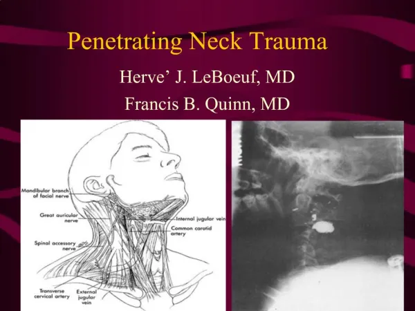
7. Anatomy First divided into zones in a paper from Monson et al Cook County Hospital 1969
Zone I clavicles to cricoid �
Zone II � cricoid to angle of the mandible
Zone III � angle of the mandible to the base of the skullFirst divided into zones in a paper from Monson et al Cook County Hospital 1969
Zone I clavicles to cricoid �
Zone II � cricoid to angle of the mandible
Zone III � angle of the mandible to the base of the skull
8. Anatomy Zone I �
inferior trachea and esophagus
vessels of the root of the neck: the brachiocephalic trunk, the subclavian arteries, the common carotid arteries, the thyrocervical trunk and the corresponding veins,
thoracic duct,
thyroid gland,
spinal cord.
Zone II �
the larynx, hypopharynx
common carotid arteries
the internal and external carotid arteries
the internal jugular veins
and cranial nerves 10, 11, and 12,
the spinal cord.
Zone III �
the pharynx
carotid arteries
the vertebral arteries
the internal jugular veinsZone I �
inferior trachea and esophagus
vessels of the root of the neck: the brachiocephalic trunk, the subclavian arteries, the common carotid arteries, the thyrocervical trunk and the corresponding veins,
thoracic duct,
thyroid gland,
spinal cord.
Zone II �
the larynx, hypopharynx
common carotid arteries
the internal and external carotid arteries
the internal jugular veins
and cranial nerves 10, 11, and 12,
the spinal cord.
Zone III �
the pharynx
carotid arteries
the vertebral arteries
the internal jugular veins
9. Incision for Neck Exploration: Zone 2 /3 injuries: �hockey stick incision.�
Open in layers
Retract the SCM laterally.
Methylene blue to aid in visualizing leaks.
Mandibulotomy or mandibular subluxation for zone 3.Zone 2 /3 injuries: �hockey stick incision.�
Open in layers
Retract the SCM laterally.
Methylene blue to aid in visualizing leaks.
Mandibulotomy or mandibular subluxation for zone 3.
10. Incisions for Neck Exploration: Zone I �
left anterior thoracotomy vs. left posterolateral thoracotomy for left zone I.
midline sternotomy with extension to the right supraclavicular region for right IZone I �
left anterior thoracotomy vs. left posterolateral thoracotomy for left zone I.
midline sternotomy with extension to the right supraclavicular region for right I
11. Incidence and Mortality Table 1 �
Mortality has decreased over the years
Most still due to exsanguination
Table 2 � McConnel paper combined data from 1963-1990 papers (2,495 pts)
Most common sight of injury is aerodigestive tract (20%)
IJ was the most commonly injured vessel followed by carotid.Table 1 �
Mortality has decreased over the years
Most still due to exsanguination
Table 2 � McConnel paper combined data from 1963-1990 papers (2,495 pts)
Most common sight of injury is aerodigestive tract (20%)
IJ was the most commonly injured vessel followed by carotid.
12. Initial Management
13. Signs of Injury:
14. Signs of Injury:
15. Management of the Stable Patient:
16. The Old Standard: Based on wartime experiences
Fogelman et al (1956) showed that immediate neck exploration led to better outcomes in study group for vascular injuries.
Led to rate of negative neck explorations in > 50%
Arteriogram slowly began to gain acceptance as screening tool before exploration, especially for zone 1 and 3 injuries (hard to detect on physical).
17. Arteriogram Zone 1 and Zone 3 vascular injuries are difficult to visualize by physical exam, making arteriogram useful in these patients.
Flint et al (1973) reported absence of P.E. findings in 32% of pts. with major zone 1 vascular injury.
Arteriogram can be accompanied by embolization.
18. A Newer Algorithm A large amount of literature accumulated showing mandatory exploration is not always necessary.A large amount of literature accumulated showing mandatory exploration is not always necessary.
19. Newer Algorithm (Mansour) 63% of the study population was in the observation group.
Entire study population had a mortality of 1.5%, similar to those in more rigorous treatment protocols.
Similar results obtained in other large studies with similar protocols (e.g. Biffi et al 1997).
Still uses the Arteriogram in asymptomatic patients with zone 1 injury.
20. Points of Controversy: Most trauma surgeons accept observation of select patients similar to the Mansour algorithm.
Study by Eddy et al questions the necessity for arteriogram / esophagoscopy in asymptomatic zone 1 injury (use of P.E. and CXR resulted in no false negatives).
Other noninvasive modalities than arteriogram exist for screening patients for vascular injury.
21. CT scan Can aid in identifying weapon trajectory and structures at risk.
Should only be used in stable patients.
Gracias et al (2001) found that use of CT scan in stable patients was able to save patients from arteriogram indicated by other protocols 50% of the time and avoid esophagoscopy in 90% of tested patients who might otherwise have undergone it. Gracias et al � metallic markers @ skin entry sites, skin violation, subQ fat stranding, soft tissue air or hematoma, vertebral fracture, contrast extravasation, missile location.Gracias et al � metallic markers @ skin entry sites, skin violation, subQ fat stranding, soft tissue air or hematoma, vertebral fracture, contrast extravasation, missile location.
22. Duplex Ultrasonography Requires the presence of reliable technician and radiologist.
A double blinded study by Ginsburg et al (1996) showed 100% true negative, 100% sensitivity in detecting arterial injury, using arteriography as the gold standard.
23. Management of Vascular Injuries: Common carotid: repair preferred over ligation in almost all cases. Saphenous vein graft may be used. Shunting is rarely necessary. Thrombectomy may be necessary.
Internal carotid: Shunting is usually necessary
Vertebral: Angiographic embolization or proximal ligation can be used if the contralateral vertebral artery is intact.
Internal Jugular: Repair vs. ligation.
24. Esophageal Injury: Best detected by combination of esophagoscopy and esophagram in symptomatic patients.
Injection of air or methylene blue in the mouth may aid in localizing injuries.
Close wounds in watertight 2 layer fashion.
Controlled fistula with T-tube or exteriorization of low non-repairable wounds
Small pharyngeal lesions above arytenoids can be treated with NPO and observation 5-7 days
All patients should be NPO for 5-7 days.
25. Laryngeal/Tracheal Injury Thorough Direct Laryngoscopy for suspicious wounds
Tracheotomy for suspected laryngeal injury
Entry incision in the cricoid cartilage, extend in the midline to the thyroid membrane, and meticulously close mucosal lacerations, using advancement flaps if necessary, or rarely, grafts. Wire vs. miniplate fixation of cartilaginous fractures.Entry incision in the cricoid cartilage, extend in the midline to the thyroid membrane, and meticulously close mucosal lacerations, using advancement flaps if necessary, or rarely, grafts. Wire vs. miniplate fixation of cartilaginous fractures.
26. Conclusions Mandatory neck exploration is no longer considered acceptable
ABC�s
Physical Exam is probably the most useful diagnostic tool.
Intervention should be directed to sites of possible injury
Non-invasive diagnostic modalities should be considered.