Download
1 / 26
270 likes | 552 Vues
Management of Penetrating Neck Trauma. Shashidhar S. Reddy, MD, MPH Shawn D. Newlands, MD, PhD. Types of Weapons. Low velocity – knives, ice picks, glass High velocity – handguns, shotguns, shrapnel. K=1/2mv^2. Guns. <. Ballistics. Ballistics. Ballistics. Anatomy. Anatomy. Zone III.

E N D
Management of Penetrating Neck Trauma Shashidhar S. Reddy, MD, MPH Shawn D. Newlands, MD, PhD
Types of Weapons • Low velocity – knives, ice picks, glass • High velocity – handguns, shotguns, shrapnel K=1/2mv^2
Guns <
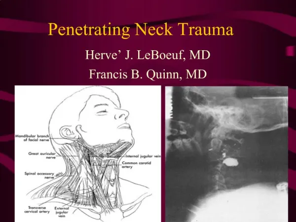
Anatomy Zone III Zone II Zone I
Initial Management Airway Intubation vs. Surgical Airway Breathing Circulation IV access, Immediate Exploration Examination Determine weapon trajectory
Signs of Injury: Shock, Profuse bleeding, Evolving stroke, Expanding hematoma, hemoptysis, hematemesis, unequal pulses, bruits or thrills Vascular
Signs of Injury: Subcutaneous emphysema, Hoarseness, Respiratory distress, Stridor Larynx/Trachea Neck pain, Blood in saliva, Fever, Odynophagia Esophagus
Management of the Stable Patient: The Old Standard: Wound Penetrates Platysma? No Yes Immediate Neck Exploration Observation/Discharge Laryngoscopy Esophagoscopy
The Old Standard: • Based on wartime experiences • Fogelman et al (1956) showed that immediate neck exploration led to better outcomes in study group for vascular injuries. • Led to rate of negative neck explorations in > 50% • Arteriogram slowly began to gain acceptance as screening tool before exploration, especially for zone 1 and 3 injuries (hard to detect on physical).
Arteriogram • Zone 1 and Zone 3 vascular injuries are difficult to visualize by physical exam, making arteriogram useful in these patients. • Flint et al (1973) reported absence of P.E. findings in 32% of pts. with major zone 1 vascular injury. • Arteriogram can be accompanied by embolization.
A Newer Algorithm Mansour et al 1991 retrospective study
Newer Algorithm (Mansour) • 63% of the study population was in the observation group. • Entire study population had a mortality of 1.5%, similar to those in more rigorous treatment protocols. • Similar results obtained in other large studies with similar protocols (e.g. Biffi et al 1997). • Still uses the Arteriogram in asymptomatic patients with zone 1 injury.
Points of Controversy: • Most trauma surgeons accept observation of select patients similar to the Mansour algorithm. • Study by Eddy et al questions the necessity for arteriogram / esophagoscopy in asymptomatic zone 1 injury (use of P.E. and CXR resulted in no false negatives). • Other noninvasive modalities than arteriogram exist for screening patients for vascular injury.
CT scan • Can aid in identifying weapon trajectory and structures at risk. • Should only be used in stable patients. • Gracias et al (2001) found that use of CT scan in stable patients was able to save patients from arteriogram indicated by other protocols 50% of the time and avoid esophagoscopy in 90% of tested patients who might otherwise have undergone it.
Duplex Ultrasonography • Requires the presence of reliable technician and radiologist. • A double blinded study by Ginsburg et al (1996) showed 100% true negative, 100% sensitivity in detecting arterial injury, using arteriography as the gold standard.
Management of Vascular Injuries: • Common carotid: repair preferred over ligation in almost all cases. Saphenous vein graft may be used. Shunting is rarely necessary. Thrombectomy may be necessary. • Internal carotid: Shunting is usually necessary • Vertebral: Angiographic embolization or proximal ligation can be used if the contralateral vertebral artery is intact. • Internal Jugular: Repair vs. ligation.
Esophageal Injury: • Best detected by combination of esophagoscopy and esophagram in symptomatic patients. • Injection of air or methylene blue in the mouth may aid in localizing injuries. • Close wounds in watertight 2 layer fashion. • Controlled fistula with T-tube or exteriorization of low non-repairable wounds • Small pharyngeal lesions above arytenoids can be treated with NPO and observation 5-7 days • All patients should be NPO for 5-7 days.
Laryngeal/Tracheal Injury • Thorough Direct Laryngoscopy for suspicious wounds • Tracheotomy for suspected laryngeal injury
Conclusions • Mandatory neck exploration is no longer considered acceptable • ABC’s • Physical Exam is probably the most useful diagnostic tool. • Intervention should be directed to sites of possible injury • Non-invasive diagnostic modalities should be considered.