Download
1 / 51
520 likes | 660 Vues
Penetrating neck trauma accounts for 5-10% of serious injuries, with a mortality rate of 5%. Learn about causes, zones, injuries, and clinical management.

E N D
frequency • 5-10% of all serious traumatic injuries • Mortality rate around 5% • Ant. And lat. Aspects are more vulnerable to trauma
Types • Blunt • Penetrating
Blunt • Causes like - Road Traffic accidents - Strangulation - Fist - Suicide
Penatrating Causes - Guns & knives 95% - Motor accidents - Sports - Household - Industrial
Guns <
Types of Injury - Penetrating • 40% do not involve important structure • Structures • major vein 15-25% • major artery 10-15% • pharynx or esophagus 5-15% • larynx or trachea 4-12% • major nerves 3-8%
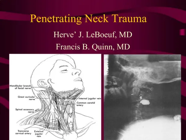
Neck Zones • Zone 1 clavicle to cricoid • Zone 2 cricoid to angle of mandible • Zone 3 above angle of mandible
Anatomy Zone III Zone II Zone I
Clinical Approach • Life Support: - Airway - Breathing - Circulation - Evaluation ( always remember cervical spine)
Airway • Debris and foreign bodies from the mouth • Ventilation through a mask • Endotracheal intubation • Surgical airway
Breathing • Air through alae nasi • Movement of chest • Auscultation for breath sounds
Studies Reviewing Airway • Eagen et al. Retrospective review of 114 pts with PNI in 8years 60% were intubated during hospital stay..........22% in E/R No complication
Madavia et al Retrospective study on 748 pts with PNI 93-96 11% emergency intubation 67% rapid sequence intubation success rate 100% 33% fibreoptic intubation,3 failed and rescued witth direct laryn.
Shearer et al retrospective study,107 pts 89-91 6% surgical airway------------------------100% 83%rapid sequence intubation----------100% 7% awake fibreoptic intubation----------98% 4% awake nasotracheal intubation-----75%
Circulation • Direct pressure for external bleeding. • Two large pore I.V access and resuscitation. • Exploration
Clinical EvaluationHistory • May be subtle , only 10% of vascular blunt trauma develop symptoms in 1st. Hour. • Zone 1 injuries may be grave with minimal presentation. • From the Patient or witnesses
History should include • a- Mechanism of injury. • b- Time elapsed from injury to presentation. • c- the patient baseline condition • d- rough determine of amount of blood • e- seat belt,location in the car and car damage • f- the weapon used :type of gun, length of knife
PresentationVascular injury • 1-Shock • 2-bleeding • 3-expanding haematoma • 4-haemoptysis • 5-haematemesis • 6-unequal pulses • 7-bruit or thrill
Presentationairway injury • 1-Subcutaineous emphysema • 2-Resp distress. • 3-Stridor and hoarsness • 4-haemoptesis
Presentation Esophagus • Neck pain • blood in saliva • Fever • Odynophagia
PresentationSpinal cord Injury • 1-complete: paraplegia or quadroplegia depending on the level. - loss of sensory and motor 2 levels below bony injury. - risk of neurogenic shock
PresentationSpinal Cord • 2-Partial injury: -central cord syndrome -ant cord syndrome -Brown-Sequard syndrome
Studies evaluating PE in Zone II • Attberry et al, prospective study on 66 pts with zone II PNI if no evidence of vascular injuryadmitted and followed up with U/S up to one year. No pts had evidence of vascular injury not anticipated by P/E.
Demetriades et al, prospective study on 82 stable PNI pts,(P/E,then admissiondoppler and angiogram) all serious injuries were detected by P/E,six were missed but didnot require intervention.
Gonzales et al, prospective study on 42 pts to compare P/E to CT scan CT scan doesnot add to the sensitivity of P/E in detecting vascular and esophageal injury.
Sekharan et al, retrospective study 145 pts (91-99) Miss rate of serious vascular or esophageal injury of 0.7%.
Management of the Stable Patient :The Old Standard: Wound penetrate platysma ? -Yes : Immediate neck exploration,. esophagoscopy, laryngoscopy. -No : Observation, closure of wound.
The Old Standard: • Based on wartime experiences • Fogelman et al(1956) showed that immediate neck exploration led to better outcomes in study group for vascular injuries. • Led to rate of negative neck explorations in > 50% • Arteriogram slowly began to gain acceptance as screening tool before exploration, especially for zone 1 and 3 injuries (hard to detect on physical).
Arteriogram • Zone 1 and Zone 3 vascular injuries are difficult to visualize by physical exam, making arteriogram useful in these patients. • Flint et al(1973) reported absence of P.E. findings in 32% of pts. with major zone 1 vascular injury. • Arteriogram can be accompanied by embolization.
Carotid Artery Dissection Internal Carotid Occlusion
Newer Algorithm • 63% of the study population was in the observation group. • Entire study population had a mortality of 1.5%, similar to those in more rigorous treatment protocols. • Similar results obtained in other large studies with similar protocols (e.g. Biffi et al1997). • Still uses the Arteriogram in asymptomatic patients with zone 1 injury.
Points of Controversy: • Most trauma surgeons accept observation of selected patients similar to the Mansour algorithm. • Study by Eddy et al questions the necessity for arteriogram / esophagoscopy in asymptomatic zone 1 injury (use of P.E. and CXR resulted in no false negatives). • Other noninvasive modalities than arteriogram exist for screening patients for vascular injury.
CT scan • Can aid in identifying weapon trajectory and structures at risk. • Should only be used in stable patients. • Gracias et al(2001) found that use of CT scan in stable patients was able to save patients from arteriogram indicated by other protocols 50% of the time and avoid esophagoscopy in 90% of tested patients who might otherwise have undergone it.
Duplex Ultrasonography • Requires the presence of reliable technician and radiologist. • A double blinded study by Ginsburg et al(1996) showed 100% true negative, 100% sensitivity in detecting arterial injury, using arteriography as the gold standard.
Management of Vascular Injuries: • Common carotid: repair preferred over ligation in almost all cases. Saphenous vein graft may be used. Shunting is rarely necessary. Thrombectomy may be necessary. • Internal carotid: Shunting is usually necessary • Vertebral: Angiographic embolization or proximal ligation can be used if the contralateral vertebral artery is intact. • Internal Jugular: Repair vs. ligation.
Treatment- Specific Injuries • Carotid injuries • 22% of penetrating cervical vascular injuries • mortality 10-20% (in-hospital) • Repair vs ligation • repair if possible in absence of neurologic deficits • prefer saphenous vein, but prosthetics ok • if internal carotid injuries, transposition of external carotid • ligation in neurologically intact for high internal carotid injury if very difficult or impossible
Esophageal Injury: • Best detected by combination of esophagoscopy and esophagram in symptomatic patients. • Injection of air or methylene blue in the mouth may aid in localizing injuries. • Close wounds in watertight 2 layer fashion. • Controlled fistula with T-tube or exteriorization of low non-repairable wounds • Small pharyngeal lesions above arytenoids can be treated with NPO and observation 5-7 days • All patients should be NPO for 5-7 days.
Tracheal Injury • Protective Tracheostomy. • If possible primary repair and muscle flap using sup. Belly of omohyoid or sternocledomasoid.