Cell Structure & Function
Cell Structure & Function. http://koning.ecsu.ctstateu.edu/cell/cell.html. Why Study Cell Biology?. The key to every biological problem must finally be sought in the cell, for every living organism is, or at some time has been, a cell. E.B. Wilson, 1925. Amoeba Proteus. Definition of Cell.


Cell Structure & Function
E N D
Presentation Transcript
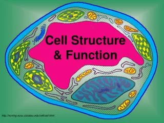
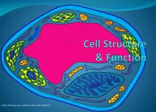
Cell Structure & Function http://koning.ecsu.ctstateu.edu/cell/cell.html
Why Study Cell Biology? The key to every biological problem must finally be sought in the cell, for every living organism is, or at some time has been, a cell. E.B. Wilson, 1925
Amoeba Proteus Definition of Cell Plant Stem Bacteria A cell is the smallest unit that is capable of performing life functions. Red Blood Cell Nerve Cell
Cells are Us Cilia on a protozoan Sperm meets egg
Red and white blood cells above vessel-forming cells. nerve cell Cells are Us A person contains about 100 trillion cells. That’s 100,000,000,000,000 or 1 x 10¹⁴ cells. There are about 200 different cell types in mammals (one of us). Cells are tiny, measuring on average about 0.002 cm (20 um) across. That’s about 1250 cells, “shoulder-to-shoulder” per inch.
Schleiden Schwann The Cell Theory The cell theory (proposed independently in 1838 and 1839) is a cornerstone of biology. All organisms are composed of one or more cells. Cells are the smallest living things. Cells arise only by division of previously existing cells. All organisms living today are descendents of an ancestral cell.
A Sense of Scale and Abundance – Bacteria on the Head of a Pin
Microscopes • Anton Von Leuwenhoek invented the first microscope in 1674 (capable of 200X magnification) Light microscopescan resolve structures that are 200nm apart. Electron microscopescan resolve structures that are 0.2nm apart.
Why Are Cells So Small? • However, as cell volume increases the surface area of the cell does not expand as quickly. • If the cell’s volume gets too large it cannot transport enough wastes out or nutrients in. • Thus, surface area limits cell volume/size.
Why Are Cells So Small? • Strategies for increasing surface area, so cell can be larger: • “Frilly” edged……. • Long and narrow….. • Round cells will always be small.
Why Are Cells So Small? • Cells need sufficient surface area to allow adequate transport of nutrients in and wastes out. • As cell volume increases, so does the need for the transporting of nutrients and wastes.
Cell Structure • All Cells have: • an outermost plasma membrane • genetic material in the form of DNA • cytoplasm with ribosomes
A prokaryotic cell A eukaryotic cell Two Fundamentally Different Types of Cells
Prokaryotic Cells Prokaryotic cells lack a membrane-bound nucleus. -genetic material is present in the nucleoid Two types of prokaryotes: -archaea -bacteria
Prokaryotic Cell Structure • Structures • Plasma membrane • Cell wall • Cytoplasm with ribosomes • Nucleoid • Capsule* • Flagella* and pili* *present in some, but not all prokaryotic cells
Prokaryotic Cells Prokaryotic cell walls -protect the cell and maintain cell shape Bacterial cell walls -may be composed of peptidoglycan -may be Gram positive or Gram negative Archaean cell walls lack peptidoglycan.
Prokaryotic Cells Flagella -present in some prokaryotic cells -used for locomotion -rotary motion propels the cell
Streptococcus pyogenes aka Necrotizing Fasciitis “flesh eating bacteria” Streptococcus pyogenes is one of the most frequent pathogens of humans. It is estimated that between 5-15% of normal individuals harbor the bacterium, usually in the respiratory tract, without signs of disease.
Staphylococcus aureus • Staphylococcus aureus • forms a fairly large yellow colony on rich medium; S. epidermidis has a relatively small white colony. • Staphylococci are facultative anaerobes that grow by aerobic respiration or by fermentation that yields principally lactic acid. • S. aureus can grow at a temperature range of 15 to 45 degrees and at NaCl concentrations as high as 15 percent. • Nearly all strains of S. aureus produce the enzyme coagulase: nearly all strains of S. epidermidis lack this enzyme. • S. aureus should always be considered a potential pathogen; most strains of S. epidermidis are nonpathogenic and may even play a protective role in humans as normal flora. .
Lactobacillus acidophilus Lactic Acid Bacteria (LAB) are Gram-positive, non-sporeforming cocci, coccobacilli or rods with a DNA base composition of less than 53mol% G+C. They generally are non respiratory and lack catalase. They ferment glucose primarily to lactic acid, or to lactic acid, CO2 and ethanol. All LAB grow anaerobically, but unlike most anaerobes, they grow in the presence of O2 as "aerotolerant anaerobes". Although they lack catalase, they possess superoxide dismutase and have alternative means to detoxify peroxide radicals, generally through peroxidase enzymes.
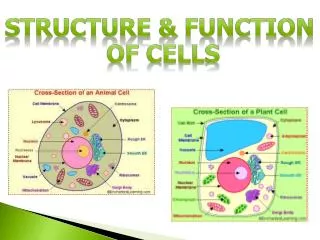
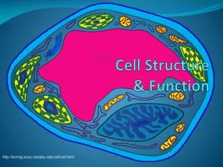
Eukaryotic Cells • Structures in all eukaryotic cells • Nucleus • Ribosomes • Endomembrane System • Endoplasmic reticulum – smooth and rough • Golgi apparatus • Vesicles • Mitochondria • Cytoskeleton
NUCLEUS CYTOSKELETON RIBOSOMES ROUGH ER MITOCHONDRION CYTOPLASM SMOOTH ER CENTRIOLES GOLGI BODY LYSOSOME PLASMA MEMBRANE VESICLE Fig. 4-15b, p.59