Unit 24: The Foot
Unit 24: The Foot. Calcaneus Talus Tarsals Navicular Cuboid Cuneiform (medial, middle, lateral) Metatarsals Phalanges. Bony Anatomy. Arches of the Foot. Metatarsal arch and transverse Medial longitudinal Lateral longitudinal. Plantar Fascia. Ligaments Subtalar ligaments

Unit 24: The Foot
E N D
Presentation Transcript
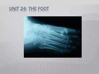
Calcaneus • Talus • Tarsals • Navicular • Cuboid • Cuneiform (medial, middle, lateral) • Metatarsals • Phalanges Bony Anatomy
Arches of the Foot • Metatarsal arch and transverse • Medial longitudinal • Lateral longitudinal
Ligaments • Subtalar ligaments • Plantar calcaneonavicular • Shock absorption • Spring ligament • Midtarsal ligaments • Anterior tarsal ligaments Soft Tissue Anatomy
Eversion –ankle Abduction – forefoot Pronation – combination movement Where are these muscles located? Muscles and Movements
Inversion – forefoot • Adduction – forefoot • Supination – combination movement • Where are these muscles located?
Phalange motion- Flexion Extension Abduction Adduction
Highly vulnerable area to variety of injuries • Injuries best prevented by selecting appropriate footwear, correcting biomechanical structural deficiencies through orthotics • Foot will adapt to training surfaces over time • Must be aware of potential difficulties associated with non-yielding and absorbent training surfaces Prevention of Foot Injuries
Athletes should be referred to qualified personnel for injury evaluation • History • Generic history questions • Questions specific to the foot • Location of pain - heel, foot, toes, arches? • Training surfaces or changes in footwear? • Changes in training, volume or type? • Does footwear increase discomfort? Foot Assessment
Observations • Does athlete favor a foot, limp, or is unable to bear weight? • Does foot color change w/weight bearing? • Is there pesplanus/cavus? • How is foot alignment? • Structural deformities? • What does wear pattern look like on the sole of the shoe? • Is the wear symmetrical?
Posterior view What do you see????
Palpation • Should assess the bony anatomy first • Checking for deformities and areas of tenderness • Assessment of soft tissue (muscles and tendons) will allow for detection of point tenderness, swelling, muscle spasm or muscle guarding • Circulation must also be monitored using the dorsal pedal pulse • Located on anterior surface of ankle and foot
Foot problems are associated with improper footwear, poor hygiene, anatomical structural deviations or abnormal stresses Sports place exceptional stress on feet ATC’s must be aware of potential problems and be capable of identifying, ameliorating or preventing them Recognition and Management of Specific Injuries
What examples of improper footware can you think of? What about flip flops? Long term problems? What should you wear after an injury? Shoe Problems
Retrocalcaneal Bursitis (Pump Bump) • Cause of Injury • Caused by inflammation of bursa beneath Achilles tendon • Result of pressure and rubbing of shoe heel counter of a shoe • Chronic condition that develops over time and may take extensive time to resolve, exostosis (pump bump) may develop • Must differentiate from Sever’s disease
Sign and Symptoms • Signs of inflammation • Tender, palpable bump on calcaneous • Pain w/palpation superior and anterior to Achilles insertion, swelling on both sides of the heel cord • Care • Routine stretching of Achilles, heel lifts to reduce stress, donut pad to reduce pressure • Select different footwear that results in increasing or decreasing height of heel counter.
Heel Bruise • Cause of Injury • Caused by sudden starts, stops, or changes of direction, irritation of fat pad • Signs of Injury • Severe pain in heel and unable to withstand stress of weight bearing • May progress to chronic inflammation of bone covering • Care • Reduce weight bearing for 24 hours, RICE and NSAID’s • Resume activity with heel cup or doughnut pad after pain has subsided (be sure to wear shock absorbent shoes)
Plantar Fasciitis • Cause of Condition • Increased stress on fascia • Change from rigid supportive footwear to flexible footwear • Poor running technique • Leg length discrepancy, excessive pronation, inflexible longitudinal arch, tight gastroc-soleus complex • Running on soft surfaces, shoes with poor support • Sign and Symptoms • Pain in anterior medial heel, along medial longitudinal arch • Increased pain in morning, loosens after first few steps • Increased pain with forefoot dorsiflexion
Care • Extended treatment (8-12 weeks) is required • Orthotic therapy is very useful (soft orthotic with deep heel cup) • Simple arch taping, use of a night splint to stretch • Vigorous heel cord stretching and exercises that increase great toe dorsiflexion • NSAID’s and occasionally steroidal injection
Metatarsal Fractures • Cause of Injury • Direct force or by placing torsional/twisting stresses on bone • Signs of Injury • Difficult to distinguish fracture from sprain in this case • Generally present with swelling, pain, point tenderness and possible deformity • X-ray will be necessary to distinguish fx from sprain • Care • Symptomatic • RICE for swelling • Short leg walking cast once swelling subsides (3-6 weeks)
Jones Fracture • Cause of Injury • Fracture of metatarsal caused by inversion or high velocity rotational forces • Most common = base of 5th metatarsal • Sign of Injury • Immediate swelling, pain over 5th metatarsal • May feel a “pop” • High nonunion rate and course of healing is unpredictable • Care • Generally requires 6-8 weeks non-weight bearing with short leg cast if non-displaced • If nonunion occurs, internal fixation may be required
Metatarsal Stress Fractures • Cause of Injury • 2nd metatarsal fracture (March fracture) • Change in running pattern, mileage, hills, or hard surfaces • Often the result of structural deformities of the foot or training errors (terrain, footwear, surfaces) • Often associated with Morton’s toe • Signs of Injury • Pain and tenderness along second metatarsal • Pain with running and walking • Continued pain/aching when non-weight bearing
Care • Determine cause of injury • Generally good success with modified rest and training modifications (pool running, stationary bike) for 2-4 weeks • Return to running should be gradual over a 2-3 week period with appropriate shoes
Metatarsal Arch Strain • Cause of Injury • Hypermobility of metatarsals caused by laxity in ligaments – results in excessive splay of foot • Will appear to have fallen arch • Signs of Injury • Pain or cramping in metatarsal region • Point tenderness (metatarsalgia), weakness • Heavy callus may form in area of pain • Care • Pad to elevate metatarsals just behind ball of foot • Strengthening of foot muscles and heel cord stretching
Longitudinal Arch Strain • Cause of Injury • Result of increased stress on arch of foot • Flattening of foot during midsupport phase causing strain on arch (appear suddenly or develop slowly) • Sign of Injury • Pain with running and jumping, usually below posterior tibialis tendon, accompanied by pain and swelling • May also be associated with sprained calcaneonavicular ligament and flexor hallucislongus strain • Care • Immediate care, RICE, reduction of weight bearing • Weight bearing must be pain free • Arch taping may be used to allow pain free walking
Fractures and Dislocations of the Phalanges • Cause of Injury • Kicking un-yielding object, stubbing toe, being stepped on • Signs of Injury • Immediate and intense pain • Swelling and discoloration • Obvious deformity with dislocation • Care • Dislocations should be reduced by a physician • Casting may occur with great toe or stiff-soled shoe • Buddy taping is generally sufficient • Shoe with larger toe box may be necessary
Bunion (HalluxValgus Deformity) • Cause of Injury • Exostosis of 1st metatarsal head; associated with forefoot varus; shoes that are too narrow, pointed, or short • Bursa becomes inflamed and thickens, enlarging joint, and causing lateral malalignment of great toe • Sign of Injury • Tenderness, swelling, and enlargement of joint initially • As inflammation continues, angulation increases causing painful ambulation
Care • Wear correctly fitting shoes, appropriate orthotics, pad over 1st metatarsal head, tape splint between 1st and 2nd toe • Surgery may be required during later stages of condition
Morton’s Neuroma • Cause of Condition • Thickening of nerve sheath (common plantar nerve) at point where nerve divides into digital branches • Commonly occurs between 3rd and 4th met heads where medial and lateral plantar nerves come together • Signs of Condition • Burning paresthesia and severe intermittent pain in forefoot • Pain relieved with non-weight bearing • Toe hyperextension increases symptoms
Care • Teardrop pad can be placed between met heads to increase space, decreasing pressure on neuroma • Shoes with wider toe box would be appropriate
Turf Toe • Cause of Injury • Hyperextension injury resulting in sprain of 1st metatarsophalangeal joint • May be the result of single or repetitive trauma • Signs and Symptoms • Pain and swelling which increases during push-off in walking, running, and jumping • Care • Increase rigidity of forefoot region in shoe • Taping the toe to prevent dorsiflexion • Rest and discourage activity until pain free • 3-4 weeks may be required for pain to subside
Calluses • Cause of Condition • Develop from friction – may be painful as fatty layer loses elasticity and cushioning effect • May be vulnerable to tears and cracks and possible blister development underneath • Care • Emery callus file may be necessary • Massaging with small amounts of lotion may be helpful • Sanding or pumicing – care must be exercised • Can be prevented • Shoes that fit appropriately are recommended • Wear at least one layer of socks • Apply petroleum jelly to reduce friction