Chapter 42
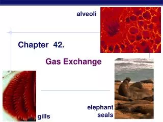
Chapter 42. Circulation and Gas Exchange. Concept 42.5: Gas exchange occurs across specialized respiratory surfaces. Gas exchange supplies oxygen for cellular respiration and disposes of carbon dioxide. Partial Pressure Gradients in Gas Exchange.

Chapter 42
E N D
Presentation Transcript
Chapter 42 Circulation and Gas Exchange
Concept 42.5: Gas exchange occurs across specialized respiratory surfaces • Gas exchange supplies oxygen for cellular respiration and disposes of carbon dioxide
Partial Pressure Gradients in Gas Exchange • Gases diffuse down pressure gradients in the lungs and other organs as a result of differences in partial pressure • Partial pressure is the pressure exerted by a particular gas in a mixture of gases
A gas diffuses from a region of higher partial pressure to a region of lower partial pressure • In the lungs and tissues, O2 and CO2 diffuse from where their partial pressures are higher to where they are lower
Respiratory Media • Animals can use air or water as a source of O2, or respiratory medium • In a given volume, there is less O2 available in water than in air • Obtaining O2 from water requires greater efficiency than air breathing
Respiratory Surfaces • Animals require large, moist respiratory surfaces for exchange of gases between their cells and the respiratory medium, either air or water • Gas exchange across respiratory surfaces takes place by diffusion • Respiratory surfaces vary by animal and can include the outer surface, skin, gills, tracheae, and lungs
Gills in Aquatic Animals • Gills are outfoldings of the body that create a large surface area for gas exchange
Fig. 42-21 Coelom Gills Gills Tube foot Parapodium (functions as gill) (a) Marine worm (c) Sea star (b) Crayfish
Fig. 42-21a Parapodium (functions as gill) (a) Marine worm
Fig. 42-21b Gills (b) Crayfish
Fig. 42-21c Coelom Gills Tube foot (c) Sea star
Ventilation moves the respiratory medium over the respiratory surface • Aquatic animals move through water or move water over their gills for ventilation • Fish gills use a countercurrent exchange system, where blood flows in the opposite direction to water passing over the gills; blood is always less saturated with O2 than the water it meets
Fig. 42-22 Fluid flow through gill filament Oxygen-poor blood Anatomy of gills Oxygen-rich blood Gill arch Lamella Gill arch Gill filament organization Blood vessels Water flow Operculum Water flow between lamellae Blood flow through capillaries in lamella Countercurrent exchange PO2 (mm Hg) in water 150 120 90 60 30 Gill filaments Net diffu- sion of O2 from water to blood 110 80 50 20 140 PO2 (mm Hg) in blood
Tracheal Systems in Insects • The tracheal system of insects consists of tiny branching tubes that penetrate the body • The tracheal tubes supply O2 directly to body cells • The respiratory and circulatory systems are separate • Larger insects must ventilate their tracheal system to meet O2 demands
Fig. 42-23 Air sacs Tracheae External opening Tracheoles Mitochondria Muscle fiber Body cell Air sac Tracheole Trachea Body wall Air 2.5 µm
Lungs • Lungs are an infolding of the body surface • The circulatory system (open or closed) transports gases between the lungs and the rest of the body • The size and complexity of lungs correlate with an animal’s metabolic rate
Mammalian Respiratory Systems: A Closer Look • A system of branching ducts conveys air to the lungs • Air inhaled through the nostrils passes through the pharynx via the larynx, trachea, bronchi, bronchioles, and alveoli, where gas exchange occurs • Exhaled air passes over the vocal cords to create sounds • Secretions called surfactants coat the surface of the alveoli
Fig. 42-24 Branch of pulmonary vein (oxygen-rich blood) Branch of pulmonary artery (oxygen-poor blood) Terminal bronchiole Nasal cavity Pharynx Larynx Alveoli (Esophagus) Left lung Trachea Right lung Bronchus Bronchiole Diaphragm Heart SEM Colorized SEM 50 µm 50 µm
Concept 42.6: Breathing ventilates the lungs • The process that ventilates the lungs is breathing, the alternate inhalation and exhalation of air
How an Amphibian Breathes • An amphibian such as a frog ventilates its lungs by positive pressure breathing, which forces air down the trachea
How a Mammal Breathes • Mammals ventilate their lungs by negative pressure breathing, which pulls air into the lungs • Lung volume increases as the rib muscles and diaphragm contract • The tidal volume is the volume of air inhaled with each breath
The maximum tidal volume is the vital capacity • After exhalation, a residual volume of air remains in the lungs
Fig. 42-25 Rib cage expands as rib muscles contract Rib cage gets smaller as rib muscles relax Air inhaled Air exhaled Lung Diaphragm INHALATION Diaphragm contracts (moves down) EXHALATION Diaphragm relaxes (moves up)
How a Bird Breathes • Birds have eight or nine air sacs that function as bellows that keep air flowing through the lungs • Air passes through the lungs in one direction only • Every exhalation completely renews the air in the lungs
Fig. 42-26 Air Air Anterior air sacs Trachea Posterior air sacs Lungs Lungs Air tubes (parabronchi) in lung 1 mm EXHALATION Air sacs empty; lungs fill INHALATION Air sacs fill
Control of Breathing in Humans • In humans, the main breathing control centers are in two regions of the brain, the medulla oblongata and the pons • The medulla regulates the rate and depth of breathing in response to pH changes in the cerebrospinal fluid • The medulla adjusts breathing rate and depth to match metabolic demands • The pons regulates the tempo
Sensors in the aorta and carotid arteries monitor O2 and CO2 concentrations in the blood • These sensors exert secondary control over breathing
Fig. 42-27 Cerebrospinal fluid Pons Breathing control centers Medulla oblongata Carotid arteries Aorta Diaphragm Rib muscles
Concept 42.7: Adaptations for gas exchange include pigments that bind and transport gases • The metabolic demands of many organisms require that the blood transport large quantities of O2 and CO2
Coordination of Circulation and Gas Exchange • Blood arriving in the lungs has a low partial pressure of O2 and a high partial pressure of CO2 relative to air in the alveoli • In the alveoli, O2 diffuses into the blood and CO2 diffuses into the air • In tissue capillaries, partial pressure gradients favor diffusion of O2 into the interstitial fluids and CO2 into the blood
Fig. 42-28 Alveolus Alveolus PCO2 = 40 mm Hg PO2 = 100 mm Hg PO2 = 40 PCO2 = 46 PCO2 = 40 PO2 = 100 Circulatory system Circulatory system PO2 = 40 PO2 = 100 PCO2 = 40 PCO2 = 46 PO2 ≤ 40 mm Hg PCO2 ≥ 46 mm Hg Body tissue Body tissue (a) Oxygen (b) Carbon dioxide
Respiratory Pigments • Respiratory pigments, proteins that transport oxygen, greatly increase the amount of oxygen that blood can carry • Arthropods and many molluscs have hemocyanin with copper as the oxygen-binding component • Most vertebrates and some invertebrates use hemoglobin contained within erythrocytes
Hemoglobin • A single hemoglobin molecule can carry four molecules of O2 • The hemoglobin dissociation curveshows that a small change in the partial pressure of oxygen can result in a large change in delivery of O2 • CO2 produced during cellular respiration lowers blood pH and decreases the affinity of hemoglobin for O2; this is called the Bohr shift
Fig. 42-UN1 Chains Iron Heme Chains Hemoglobin
Fig. 42-29 100 O2 unloaded to tissues at rest 80 O2 unloaded to tissues during exercise 60 O2 saturation of hemoglobin (%) 40 20 0 0 20 40 60 80 100 Tissues during exercise Tissues at rest Lungs PO2 (mm Hg) (a) PO2 and hemoglobin dissociation at pH 7.4 100 pH 7.4 80 pH 7.2 Hemoglobin retains less O2 at lower pH (higher CO2 concentration) 60 O2 saturation of hemoglobin (%) 40 20 0 0 20 40 60 80 100 PO2 (mm Hg) (b) pH and hemoglobin dissociation
Fig. 42-29a 100 O2 unloaded to tissues at rest 80 O2 unloaded to tissues during exercise 60 O2 saturation of hemoglobin (%) 40 20 0 20 40 80 100 0 60 Tissues during exercise Tissues at rest Lungs PO2 (mm Hg) (a) PO2 and hemoglobin dissociation at pH 7.4
Fig. 42-29b 100 pH 7.4 80 pH 7.2 Hemoglobin retains less O2 at lower pH (higher CO2 concentration) 60 O2 saturation of hemoglobin (%) 40 20 0 0 20 40 60 80 100 PO2 (mm Hg) (b) pH and hemoglobin dissociation
Carbon Dioxide Transport • Hemoglobin also helps transport CO2 and assists in buffering • CO2 from respiring cells diffuses into the blood and is transported either in blood plasma, bound to hemoglobin, or as bicarbonate ions (HCO3–) Animation: O2 from Blood to Tissues Animation: CO2 from Tissues to Blood Animation: CO2 from Blood to Lungs Animation: O2 from Lungs to Blood
Fig. 42-30 Body tissue CO2 transport from tissues CO2 produced Interstitial fluid CO2 CO2 Capillary wall Plasma within capillary CO2 H2O Hemoglobin picks up CO2 and H+ Red blood cell H2CO3 Hb Carbonic acid HCO3– Bicarbonate H+ + HCO3– To lungs CO2 transport to lungs HCO3– HCO3– H+ + Hemoglobin releases CO2 and H+ Hb H2CO3 H2O CO2 CO2 CO2 CO2 Alveolar space in lung
Fig. 42-30a Body tissue CO2 transport from tissues CO2 produced Interstitial fluid CO2 Capillary wall CO2 Plasma within capillary CO2 H2O Hemoglobin picks up CO2 and H+ Red blood cell H2CO3 Hb Carbonic acid HCO3– Bicarbonate H+ + HCO3– To lungs
Fig. 42-30b CO2 transport to lungs HCO3– HCO3– H+ + Hemoglobin releases CO2 and H+ Hb H2CO3 H2O CO2 Plasma within lung capillary CO2 CO2 CO2 Alveolar space in lung
Elite Animal Athletes • Migratory and diving mammals have evolutionary adaptations that allow them to perform extraordinary feats
The Ultimate Endurance Runner • The extreme O2 consumption of the antelope-like pronghorn underlies its ability to run at high speed over long distances
Fig. 42-31 RESULTS Goat Pronghorn 100 90 80 70 60 Relative values (%) 50 40 30 20 10 0 VO2 max Lung capacity Muscle mass Mitochon- drial volume Cardiac output
Diving Mammals • Deep-diving air breathers stockpile O2 and deplete it slowly • Weddell seals have a high blood to body volume ratio and can store oxygen in their muscles in myoglobin proteins
Fig. 42-UN2 Inhaled air Exhaled air Alveolar spaces Alveolar epithelial cells CO2 O2 CO2 O2 Alveolar capillaries of lung Pulmonary veins Pulmonary arteries Systemic veins Systemic arteries Heart Systemic capillaries O2 CO2 O2 CO2 Body tissue