Download
1 / 21
210 likes | 442 Vues
Diseases of the gastrointestinal tract and liver. Lecture 30 Tuesday, March 13, 2007 Refs. Basic Pathology Chapters 15 and 16 Wheater’s Basic Histopathology Chapters 13 and 14. Treatment of ulcers. Biopsy to determine cause For H. pylori infection:

E N D
Diseases of the gastrointestinal tract and liver Lecture 30 Tuesday, March 13, 2007 Refs. Basic Pathology Chapters 15 and 16 Wheater’s Basic Histopathology Chapters 13 and 14
Treatment of ulcers • Biopsy to determine cause • For H. pylori infection: • Triple therapy of antibiotics and proton pump inhibitor • Patient compliance issues • For NSAIDs: • Discontinue use • Switch to a COX-2 inhibitor • For carcinoma: • Surgery
Inhibition of gastric acid secretion • Proton pump inhibitors • Omeprazole • Histamine-2 receptor inhibitors
Malignant gastric neoplasia • Most common- adenocarcinoma • Prognosis is good if tumor has not invaded muscle wall • Intestinal type • Gland formation, moderate to well-differentiated • Diffuse • Sheets of neoplastic cells, signet ring cells • Second most common(~5%)-lymphoma • Lymphoepithelial lesion- infiltration of epithelium • MALToma may regress after eradication of H. pylori • Rare-carcinoid
Carcinoid • A neoplasm of neuroendocrine cells • Besides stomach, can also occur in the small intestine, pancreas, biliary tract, and lung • May secrete hormone • Cells appear benign -uniform,small round cells • Locally invasive • Potential to metastasize
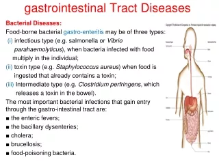
Diseases of the small intestine • Enteritis (table 15-7) • Viral or bacterial, usually self-limiting • If diarrhea causes dehydration, can be fatal • Malabsorption syndrome- If more than 50% of small intestine is removed, malnutrition occurs. • Celiac disease • Synonyms: nontropical sprue, celiac sprue, gluten-sensitive enteropathy • Gluten sensitivity, T cell mediated • Genetic predisposition • Villi undergo atrophy, can be restored on gluten-free diet
Diseases of the small intestine con’t • Protozoa- Giardiasis • Protozoan flagellate can cause blunting of villi and malabsorptive diarrhea • Contaminated water, fecal-oral • Whipple disease is a systemic disease that can cause malabsorption • Macrophages in lamina propria are filled with bacilli • Tropheryma whippelii gram-positive culture resistant actinomycete • Usually responds to antibiotic treatment • Crohn’s disease • Inflammatory disease of GI tract- segmental lesions usually in small intestine, often in colon, sometimes in stomach. • Idiopathic, possibly immune-mediated systemic inflammatory disease • Patchy distribution of lesions. • Lesions have deep ulcers and noncaseating granulomas.
Crohn’s diseaseSmall intestine with ulcer, edema and inflammation of the submucosa including granulomas. Whp 13.16
Diseases of the colon • Ulcerative colitis • Idiopathic ulcerative disease of rectum and colon • Does not involve small intestine • Diverticulitis • Neoplasia-adenomatous polyps to adenocarcinoma • Most polyps are benign • Adenocarcinoma is a frequent cause of death • Several familial syndromes of high risk of cancer
Inflammatory bowel disease (IBD) • Crohn’s disease and ulcerative colitis • Both are idiopathic. • Both have systemic inflammatory manifestations. • Familial aggregations, but no clear inheritance pattern. • Associated with different HLA antigens. • Probably defective regulation of immune response. • Intermittent attacks can be precipitated by physical and emotional stress. • Both respond to immunosuppression
Histologic differences in IBD • Crohn’s disease: deep ulcers, granulomas, transmural inflammation, mural thickening. • Ulcerative colitis: shallow ulcers, pseudopolyps (remaining mucosa between areas of ulceration), inflammation is more superficial, no granulomas. • See Robbins schematic figure 15-28
Diverticulitis • Inflammation of diverticula • A diverticulum is an outpouching of the colonic mucosa through the circular muscular layer where the longitudinal layer is thin (between tenia coli). • Common in older adults- prevalence is ~50% in people over 60 western industrialized countries. • When the neck of the diverticulum becomes obstructed, diverticulitis may develop. • Possible sequelae: perforation, peritonitis, hemorrhage, abscess and fistula formation.
Appendicitis • Common problem (10% of people) • Any age but peak incidence in 20s and 30s • Acute inflammation of vermiform appendix • May be associated with obstruction of its lumen • Pain, nausea, and vomiting • Surgical emergency • Perforation • Can cause fatal peritonitis
Physiologic factors in the susceptibility of liver to disease • Liver has high metabolic requirements. • Decreased oxygen causes damage. • Exposed to most of the blood draining from the intestinal tract including any absorbed toxins, microbes, metastatic tumor cells, etc. • Vasculature is not designed to withstand high pressure. • Right heart failure damages liver. • Anatomy of bile duct and pancreatic duct. • Pancreatitis can cause cholestasis.
Challenges in diagnosing liver disease • Enormous functional reserve masks early liver damage. • Clinical signs of liver disease may reflect the many functions it performs. • Many clinical signs of liver disease can be caused by diseases of other systems. • Edema, polyuria • Many enzyme tests are not specific because the enzymes are not unique to liver. • LDH, AST (SGOT) aspartate aminotransferase, ALT (SGPT) alanine aminotransferase • ALT is more specific for liver but does not mean necrosis • Liver has a limited number of responses to injury.
Morphologic responses to injury • 1.Inflammation = hepatitis • 2. Degeneration of hepatocytes • Steatosis (microvesicular or macrovesicular) • Iron, copper, or bile accumulation in hepatocyte • 3. Necrosis and apoptosis • Coagulative necrosis- often due to ischemia • Can be caused by toxins, viruses, etc • Councilman bodies represent apoptotic cells • Submassive necrosis- almost whole lobule is necrotic • 4. Fibrosis- generally irreversible • 5. Cirrhosis • Subdivided into nodules of regenerating hepatocytes
Outcomes of liver injury • Regeneration occurs in most diseases. • It can be almost perfect if the CT framework is intact. • Nodules of new hepatocytes with abnormal architecture • Fibrosis- collagen formed. • Affects hepatic blood flow and bile flow • Cirrhosis = interlacing fibrous bands of CT dividing the parenchyma into nodules • End stage form of liver disease.