Download

1 / 71
740 likes | 1.19k Vues
Anatomy of the peritoneum. SUAT CAN ULUKENT , M.D. PHD GENERAL SURGEON - ANATOMIST. Abdominal Cavity-Serous Membranes. The fist represents an organ and the baloon represents peritoneum.

E N D
Anatomy of the peritoneum SUAT CAN ULUKENT, M.D. PHD GENERAL SURGEON - ANATOMIST
Abdominal Cavity-Serous Membranes • The fist represents an organ and the baloon represents peritoneum. • The inner balloon wall in contact with the fist represents the visceralserous membrane (visceral peritoneum)covering the organ. • The outer part of the balloon wall represents the parietalserous membrane (parietal peritoneum). • The cavity or space betweenthe visceral and parietal serous membranes (peritoneal cavity) is normallyfilled with a thin, lubricating film of serous fluid. • This fluid reduces friction as organs rub against the body wall or against anotherorgan.
Etimology for Peritoneum • Peritoneum is derived from Greek via Latin. Peri- means around, while -ton- refers to stretching. Thus, peritoneum means stretched around or stretched over.
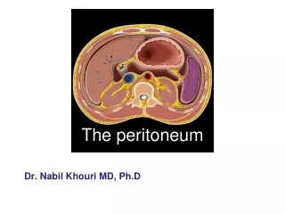
The peritoneum is in the form of a closed sac invaginated by a number of viscera • Parietal peritoneum – outer layer-lines the walls of the abdominal and pelvic cavities • Visceral peritoneum – inner layer-covers the organs • Peritoneal cavity –the potential space between the parietal and visceral layer of peritoneum • In the male, it is a closed sac, but in the female, there is a communication with the exterior through the uterine tubes, the uterus, and the vagina
Parietal peritoneum • Embryologically, it is derived from the somatopleural layer of the lateral plate mesoderm. • Blood supply & nerve supply are the same as those of the overlying body wall, except the central part of the diaphragmatic peritoneum is supplied by the phrenic nerves; peripherally, the diaphragmatic peritoneum is supplied by the lower six thoracic nerves. The parietal peritoneum in the pelvis is mainly supplied by the obturator nerve, a branch of the lumbar plexus.
Parietal peritoneum It is pain sensitive because of the somatic innervation. The parietal peritoneum is sensitive to pain, temperature, touch, and pressure. It can be easily stripped as it is loosely attached to the walls by extra-peritoneal connective tissue (fasia- fatty tissue).
Visceral peritoneum • Embryologically it is derived from the splanchno-pleural layer of the lateral plate mesoderm. • Blood supply & nerve supply are the same as those of the underlying viscera. • It is pain insensitive because of the autonomic innervation. • It is firmly adherent to the viscera & cannot be stripped; forms part & parcel of the viscera.
Visceral peritoneum The visceral peritoneum is sensitive only to stretch and tearing and is not sensitive to touch, pressure, or temperature. It is supplied by autonomic afferent nerves that supply the viscera or are traveling in the mesenteries. Overdistentionof a viscus leads to the sensation of pain. The mesenteries of the small and large intestines are sensitive to mechanical stretching.
The relationship between viscera and peritoneum • Many organs in the abdomen are suspended by folds of peritoneum. Such organs are mobile/ intraperitoneal. The degree & direction of mobility are governed by the size & direction of peritoneal fold. • Other organs are fixed & immobile. They rest directly on the posterior abdominal wall, & may be covered by peritoneum on one side. Such organs are said to be retroperitoneal.
Intraperitoneal viscera Interperitoneal viscera Retroperitoneal viscera Intraperitoneal viscera-viscera completely surrounded by peritoneum. stomach, superior part of duodenum, jejunum, ileum, cecum, vermiform appendix, transverse and sigmoid colons, spleen and ovary Interperitoneal viscera-most part of viscera surrounded by peritoneum. liver, gallbladder, ascending and descending colon, upper part of rectum, urinary bladder and uterus Retroperitoneal viscera-lie on the posterior abdominal wall & covered by peritoneum on anterior surface only kidney, suprarenal gland, pancreas, descending and horizontal parts of duodenum, middle and lower parts of rectum, and ureter
Classifications of Organs in relation to Peritoneum • A useful mnemonic to aid recollection of the abdominal retroperitoneal viscera is SAD PUCKER: • S = Suprarenal glands (aka the adrenal glands) • A = Aorta/IVC • D = Duodenum (2nd and 3rd segments [some cases the 4th segment] ) • P = Pancreas (tail is intraperitoneal) • U = Ureters • C = Colon (only the ascending and descendingbranches) • K = Kidneys • E = Esophagus • R = Rectum
Peritoneal folds are given various names! • Prefix ‘mes’ or ‘meso’ (in the middle of), followed by the name of the organ • Small intestine(enteron)- mesentery • Colon- mesocolon • Large folds attached to the stomach are called omenta (omentum=cover) • Double-layered folds of peritoneum connect organs to the abdominal wall or to each other- called ligaments • Gastrosplenic ligament
Peritoneal folds • They are best understood by recapitulating the embryology of the gut • Developing gut- divisible into 3 parts foregut, midgut & hindgut • Each part has its own artery which is a ventral branch of the abdominal aorta • Coeliac- foregut, SMA- midgut, IMA-hindgut
Foregut- esophagus, stomach, upper part of duodenum (till CBD opening in D2) • Midgut- rest of duodenum, jejunum, ileum, appendix, caecum, ascending colon, right 2/3rd of transverse colon • Hindgut- left1/3rd of transverse colon, descending colon, sigmoid colon, rectum, upper part of anal canal upto pectinate line
The abdominal part of the foregut is suspended by mesenteries both ventrally & dorsally Dorsal mesogastrium Ventral mesogastrium
….. • The ventral mesogastrium becomes divided by the developing liver into a ventral part & a dorsal part.
Fate of Ventral mesogastrium • The ventral part forms the ligaments of the liver, namely: • falciform ligament • triangular ligaments • coronary ligaments • The dorsal part forms the lesser omentum
Fate of dorsal mesogastrium • Caudal part becomes greatly elongated to form the greater omentum • Cranial part forms the gastrophrenic ligament
Fate of dorsal mesogastrium • The spleen develops in relation to the cranial part & divides it into dorsal & ventral parts. • Dorsal part forms the lienorenal ligament. • Ventral part forms the gastrosplenic ligament.
The midgut & hindgut have only a dorsal mesentery which forms the mesentery of the jejunum and ileum, the meso-appendix, transverse mesocolon & the sigmoid mesocolon. • The mesenteries of the duodenum, ascending colon, descending colon & the rectum are lost during development.
The peritoneal cavity is divided broadly into two parts • Main larger part is known as the greater sac • Smaller part situated behind the stomach, lesser omentum & liver is known as the lesser sac • Two sacs communicate through the epiploic foramen(of Winslow)
Sex differences Male • Peritoneum is a closed sac lined by mesothelium (flattened epithelium) Female • peritoneal cavity communicates with the exterior thro the uterine tubes
Structures which are formed by peritoneum Omentum fold of peritoneum that extends from stomach to adjacent organs Greater omentum
Greater omentum four-layered fold of peritoneum, the anterior two layers descend from the greater curvature of stomach and superior part of duodenum and hangs down like an apron in front of coils of small intestine, and then turns upward and attaches to the transverse colon.
Lesser omentum two layered fold of peritoneum which extends from porta hepatis to lesser curvature of stomach and superior part of duodenum • Hepatogastric ligament extends from porta hepatis to lesser curvature of stomach • Hepatoduodenal ligament Extends from porta hepatis to superior part of duodenum Contains common bile duct, proper hepatic a. and hepatic portal v. Lesser omentum
Omental foramen • Behind the right border of hepatoduodenal ligament • Superior-caudate lobe of liver • Inferior-superior part of duodenum • Anterior-hepatodudenal ligament • Posterior-peritoneum covering the inferior vena cava
Omental bursa Position-situated behind the lesser omentum and stomach Walls • Superior-peritoneum which covers the caudate lobe of liver and diaphragm • Anterior-formed by lesser omentum, peritoneum of posterior wall of stomach, and anterior two layers of greater omentum • Inferior-conjunctive area of anterior and posterior two layers of greater omentum • Posterior-formed by posterior two layers of greater omentum, transverse colon and transverse mesocolon, peritoneum covering pancreas, left kidney and suprarenal gland
Left-formed by the spleen, gastrosplenic ligamentand splenorenal ligament • Right-formed by omental foramen The Omental bursa (lesser sac) communicates with the greater sac through the omental foramen.
Mesenteries or mesocolons-two-layered fold of peritoneum that attach part of the intestines to the posterior abdominal wall
Mesenteries: result from the invagination of "intraperitoneal" organs into the sac. The mesenteries connect viscera to the posterior abdominal wall and are VERY important in that they conduct blood vessels and nerves.
Mesentery suspends the small intestine from the posterior abdominal wall • Broad and a fan-shaped • Consists of two peritoneal layers • Intestinal border-folded, 7 m long • Root of mesentery • 15 cm long • Directed obliquely from left side of L2 to in front of right sacroiliac joint
Mesoappendix • Triangular mesentery-extends from terminal part of ileum to appendix • Appendicular artery runs in free margin of the mesoappendix
Transverse mesocolona double fold of peritoneum which connects the transverse colon to the posterior abdominal wall Sigmoid mesocolon inverted V-shaped, with apex located in front of left ureter and division of common iliac artery
Ligaments -two-layered folds of peritoneum that attached the lesser mobile solid viscera to the abdominal wall Ligaments of liver • Falciform ligament of liver • Consists of double peritoneal layer • Extends from anterior abdominal wall (umbilicus) to liver • Free border of ligament site of ligamentum teres
Coronary ligament the area between upper and lower parts of the coronary ligament is the bare area of liver, this area is devoid of peritoneum and lies in contact with the diaphragm • Left and right triangular ligaments formed by right extremity of coronary ligament and left leaf of falciform ligament, respectively
Hepatogastric ligament • Hepatoduodenal ligament • Ligamentum teres hepatis
Ligaments of spleen • Gastrosplenic ligament-a double layer of peritoneum that connects the fundus of stomach to hilum of spleen. In this double layer of peritoneum are the short gastric and left gastroepiploic vessels • Splenorenal ligament-extends between the hilum of spleen and anterior aspect of left kidney. The splenic vessels lies within this ligament, as well as the tail of pancreas • Phrenicosplenic ligament • Splenocolic ligament
Ligaments of stomach • Hepatogastric ligament • Gastrosplenic ligament • Gastrophrenic ligament • Gastrocolic ligament • Gastropancreatic ligament
Lig.falciforme Lig. teres lig.hepatogastricum lig.hepatoduodenale lig.gastrophrenicum lig.gastrolienale lig.lienorenale lig.phrenicocolicum lig.gastrocolicum Omentum majus