Understanding the Gross Anatomy of the Heart and Its Circulatory Functions
This exercise explores the gross anatomy of the heart, situated within the mediastinum and encased by the pericardial cavity. It details the structure of the pericardium, divides the heart's function into the right and left sides concerning pulmonary and systemic circulation, and explains coronary circulation. Additionally, it covers the conduction system, electrical activity, and EKG waveforms, including P, QRS, and T waves. Understanding these elements is crucial for diagnosing arrhythmias and assessing heart health through hands-on laboratory exercises with sheep hearts and EKG recordings.


Understanding the Gross Anatomy of the Heart and Its Circulatory Functions
E N D
Presentation Transcript
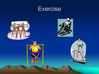
Exercise 27 Gross Anatomy of the Heart BI 232
Mediastinum • The heart and pericardial cavity are located within the mediastinum, a centrally located area within the thoracic cavity.
The Pericardium • Membranous sac • 2 parts: outer fibrous pericardium that is fused to adjacent structures and an innerserous pericardium which is a delicate serous membrane that forms a double-layered sac around the heart. • The serous pericardium consists of parietal and visceral layers.
The heart • Two-sided, double-pumping organ. • The left side controls the flow of blood to all tissues and cells in the body, where oxygen and nutrients are delivered and wastes are taken away. • The right side sends blood to the lungs, where oxygen stored in RBCs is replenished and CO2 is released
Coronary Circulation • On the posterior surface of the heart, the coronary artery branches to become the posteriorinterventricular artery • Near the apex of the heart the anterior and posterior interventricular arteries form an anastomosis (connection)
Coronary Circulation • On the posterior surface of the heart, the coronarysinus runs along the coronary sulcus and empties into the right atrium. • Great cardiac vein found in the anterior interventricularsulcus • Middle cardiac vein ascends along the posterior interventricular groove
Blood flow Through the Heart • Pulmonary Circulation- Blood from the right side of the heart flows to and from the lungs • Systemic Circulation- Blood from the left side of the heart flows from and to all body tissues.
Position of Heart • Base of the heart extends from the inferior border of the second costal cartilage on the left side to the superior border of the third costal cartilage on the right. • Apex of the heart is located in the left fifth intercostal space, left of the median plane
Heart: muscular pump • Actually 2 pumps acting in unison • Systole is contraction of heart muscle • Atrial systole • Ventricular systole • Diastole is relaxation of heart muscle • Atrial diastole • Ventricular diastole
Conduction system of the Heart (Exercise 32) • Electrical activity of the heart stimulates the heart muscle to contract. • Sinoatrial (SA) node = pacemaker of the heart • Atrioventricular (AV) node: there is a slight delay (0.10 second) • Atrioventricular bundle (bundle of His) • Right and left bundle branches • Purkinje fibers: stimulate cardiac muscle of the ventricles to contract http://youtu.be/te_SY3MeWys
Electrical activity of the Heart • Heart produces low-voltage electrochemical impulses • The average potential difference is -90 millivolts • These impulses can travel through the saline medium of the body and be picked up by sensors attached to the skin • The collective action potentials generated by the atria and ventricles depolarizing and repolarizing can be recorded using an electrocardiograph machine
P wave • The P wave indicates the depolarization of the atria just prior to the beginning of atrial contraction or systole
QRS complex (QRS interval) • Represents the depolarization of the ventricles which precedes ventricular systole.
T wave • Results from ventricular repolarization, which occurs before ventricular relaxation or diastole.
Analysis of the ECG • Irregularities in Heart rate: • Tachycardia: Above 100 in adults (in small children this may be normal) • Bradycardia: in young adults below 60 beats per minute (unless person is highly trained aerobic athlete)
Heart Blocks • PR intervals are normally about 0.16 second. • Time between the beginning of atrial depolarization and the beginning of ventricular depolarization. • Long PR might indicate a heart block (reduced conduction between atria and ventricles) • Longer than 0.2 second • Complete heart block the atria do not stimulate ventricular depolarization at all, so atria fire independently of ventricles
Heart Blocks • Normal QRS is 0.08 to 0.10 second on average. • Longer than 0.12 may indicate a right or left bundle branch block • QT interval is normally about 0.3 second • This is shorter as the heart rate increases and longer as heart rate slows.
Cardiac Arrhythmias • A major diagnostic use is to detect arrhythmias, abnormal rhythms of the heart • Atrial fibrillation: electrical activity of the heart is uncoordinated causing the upper chambers to quiver. • Ventricular fibrillation: where electrical signals in the ventricles fire in a very fast uncontrolled manner. • Premature ventricular contraction (PVC): electrical signal causes an early heartbeat that usually goes unnoticed
Today’s lab • Dissect sheep heart • ID structures on the heart models • We will also be doing EKGs (We will be using the Vernier EKGs rather than the BIOPAC machines mentioned in the lab manual) • After performing your EKG follow the instructions in your lab manual and calculate the mean electrical axis of your heart
The End The End
![[Exercise Name] Functional Exercise](https://cdn0.slideserve.com/621913/exercise-name-functional-exercise-dt.jpg)

![[Exercise Name] Functional Exercise](https://cdn1.slideserve.com/1717560/exercise-name-functional-exercise-dt.jpg)


![[Exercise Name] Functional Exercise](https://cdn3.slideserve.com/6680259/exercise-name-functional-exercise-dt.jpg)

![[Exercise Name] Tabletop Exercise](https://cdn4.slideserve.com/9191716/exercise-name-tabletop-exercise-dt.jpg)