Download

1 / 42
430 likes | 589 Vues
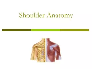
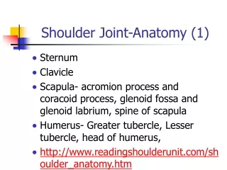
Shoulder Anatomy. Shoulder Anatomy - bones. Scapula Clavicle Humerus greater tubercle. Scapula. Supraspinous fossa. Infraspinous fossa. Subscapular fossa. Axillary border. Inferior angle. Humerus. Joints of the shoulder Sternoclavicular (SC) Joint Acromioclavicular (AC) Joint

E N D
Shoulder Anatomy - bones • Scapula • Clavicle • Humerus • greater tubercle
Scapula Supraspinous fossa Infraspinous fossa Subscapular fossa Axillary border Inferior angle
Joints of the shoulder Sternoclavicular (SC) Joint Acromioclavicular (AC) Joint Glenohumeral (GH) Joint Scapulothoracic Articulation Shoulder Anatomy
Sternoclavicular (SC) Joint • Synovial Joint - double gliding joint • side to side & front to back • Ligaments • Anterior SC • Posterior SC • Interclavicular • Costoclavicular
SC Movements • moves in many directions (but only in small amounts) • Protraction • Retraction • Elevation • Depression
SC Joint Function • Absorbs forces • Distributes forces • Allows movement of clavicle
Shoulder Anatomy - joints • Acromioclavicular Injury – “Separated Shoulder”
Acromioclavicular joint (AC) Support a. Capsule - dense but weak b. Acromioclavicular ligaments • Superior and inferior c. Coracoclavicular ligaments • Trapezoid- Lateral • Conoid -Medial
A-C Ligament: strengthens A-C joint along with fibers from trapezius Coracoclavicular Ligament: anchors Clavicle to coracoid process; strongest ligament binding the clavicle to scapula Acromioclavicular Joint
Two Parts Conoid ligament (medial) Trapezoid (lateral) A-C Separation 1-Stretch A-C 2-Complete tear of A-C; partial tear of C-C 3-Complete tear of A-C; complete tear of C-C Coracoclavicular Ligament
Shoulder Anatomy - joints • Scapulothoracic • muscular attachments only • The scapula meets the rib cage!
Scapulothoracic Articulation • Movements: • Protraction • Retraction • Elevation • Depression • Upward Rotation • Downward Rotation
Shoulder Anatomy - joints • Glenohumeral • ball and socket • lacks bony stability (small glenoid, large humerus) • provides motion • glenoid labrum (fibrocartilage) • rotator cuff (muscular stability)
Glenohumeral (GH) Joint • “True Shoulder Joint” • Synovial Joint - Ball and Socket • Glenoid fossa & labrum with Humeral Head • 3 Major Ligaments: • Coracohumeral - coracoid to greater tubercle (strong) • 3 GH bands - thickenings of articular capsule • Transverse Humeral - greater to lesser tubercle
Glenoid Cavity (shallow) Glenoid labrum (soft cartilaginous rim that adds depth and stability) Glenohumeral Stability
GH Joint Support • Capsule • Ligaments • Anterior – Glenohumeral • Superior - Coracohumeral • Coracoacromial arch • Bursae • Subacromial • Subdeltoid • Subscapular bursa
Coracohumeral ligament Glenohumeral ligaments
Anterior Capsule Posterior Capsule Glenohumeral ligaments Superior Middle Inferior Glenohumeral Stability
GH Movements • Sacrifices stability for mobility • Flexion • Extension • Abduction • Adduction • Internal Rotation • External Rotation
Upper Arm & Shoulder Muscles deltoid brachialis triceps brachii teres major teres minor
supraspinatus Rotator Cuff Muscles infraspinatus teres minor subscapularis