Transcription control in eukaryotes
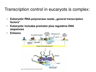
Transcription control in eukaryotes. Subjects, covered in this lecture. Overview Structural classification of eukaryotic transcription factors Transcription control mechanicms -by altered states of chromatin -through Mediator -by epigenetic mechanisms

Transcription control in eukaryotes
E N D
Presentation Transcript
Subjects, covered in this lecture • Overview • Structural classification of eukaryotic transcription factors • Transcription control mechanicms -by altered states of chromatin -through Mediator -by epigenetic mechanisms • Control of transcription factor activity • Nuclear receptors
A schematic picture of transcriptional initiation in eukaryotes
Structural classification of specific eukaryotic transcription factor domains
Homeodomains • Sequences of 60 residues that function as DNA binding domains of transcription factors • Built up from 3 helices, where helices 2 and 3 form helix-turn-helix motif similar to those in prokaryotic DNA binding proteins • First identified in Drosophila, where mutant homeodomains cause so-called homeotic transformations. Those are bizarre developmental anomalies – like legs growing from head in place of antennae.
Binding of the helix-turn-helix motif of an Antennapedia homeodomain protein in the major groove of the homeobox
DNA binding of the two domains in the POU region of the human protein Oct-1, which regulates transcription of small nuclear RNA genes and the histone H2B gene Homeodomainscan operate in tandems with similar or different DNA binding domains
Zinc fingers • The classic zinc finger motif with two histidines and two cysteines binding to the zinc ion (C2H2 type) • Other mononuclear zinc finger motifs can have three or fourcysteines. The 3D structures of those are quite different from C2H2 type – they lack the b strands.
Zinc finger proteins are multi-domain, with 1-60 tandem zinc finger DNA-binding domains and several other domains which may be responsible for dimerization, ligand or other protein binding Repression Zn Zn Zn Zn Zn Activation Example: Human GL1 protein has 5 ZNF domains and additional domains for both transcription activation and repression. Only 4 of 5 ZNFs bind to DNA
A binuclear zinc finger binding in GAL4 • Binuclear zinc finger proteins contain six Cys/His residues and two zinc ions
Dimer of GAl4 bound to UAS • Almost all contacts to DNA in Zn cluster regions are made by protein main chain atoms • Linker region determines the specificity of Zn2C6 containg proteins
Leucine zippers • Leucine zipper motif is built of two a-helices, which are kept together by hydrophobic interactions. Each seventh residue is leucine, hence the name leucine zipper • Dimer formation can be promoted by additional charge interactions
Binding of leucine zippers to DNA • Leucine zipper DNA binding proteins are homo or heterodimers • The C-terminal part of helice contains leucine zipper dimerization region, whereas N-terminal part binds to DNA and contains many basic residues
Helix-loop-helix domains • Helix-loop helix domains are somewhat similar to leucine zippers, except that a four-bundle helix motifs hold together basic DNA-binding helices
Structure of human oncogene Max is an example of combined leucine zipper – helix-loop-helix protein
Other eukaryotic DNA-binding domains Rel homology domains (NFkB, NFAT) Stat protein family DNA- binding domains with an immunoglobulin – like fold. Loops, which connect the b-strands, interact with DNA
Homo- and heterodimeric combinations of transcription factors • From three different DNA-binding protein monomers it is possible to create six different dimers with distinct binding sites
Cooperative binding of NFAT and AP1 transcription factors at IL-2 promoter
Cooperative binding of specific transcription factors can form an enhancesome Scheme for enhanceosome formation at b-interferon enhancer. Two monomeric factors IRF3 and IRF7 and two dimeric ATF-2/cJun and p50/p65 (NF-kB). HMGI is sequece-nonspecific factor, which bends DNA by binding in minor groove. It also coordinates the binding of other proteins each to other.
Molecular mechanisms of transcription activation and repression • 1. Chromatin mediated transcription control • 2. Transcription control through the Mediator • 3. Epigenetic control through DNA methylation
Background: nucleosome structure Histone monomer
Acetylation of the N-terminal sequence of histone H3 • ARTKQTARKSTGGKAPRKQL HAT (histone acetyltransferase) Histone DNA O -O-P-O O O -O-P-O O O NH3+ NH-C CH3 Unacetylated Acetylated HDAC (histone deacetylase)
URS1 – Upstream regulatory sequence DBD and RD – DNA binding and repressor domains of UME6 repressor RPD3 – yeast histone deacetylase (component of deacetylation complex) Sin3 – RD binding component of deacetylation complex
UAS – upstream activation sequence AD –activation domain of Dcn4 transcription activator Gcn5 – histone acetylase subunit of acetylation complex
Chromatin remodelling factors • Chromatin remodellig factors are multiprotein complexes with some subunits showing helicase activity • Chromatin remodelling complex SWI/SNF transiently dissociates DNA from the surface of nucleosomes, decondensing the chromatin and making the DNA more accessible to transcription factors • The activity of complex may result also in transcription repression, probably by exposing the histone tails to deacetylases or by assisting in folding of chromatin into higher-order structures
Structure of yeast and human mediator complexes • Composed of ~20 subunits which are arranged in modules • Some subunits interact with RNA Pol II, others – with activators • One subunit has histone acetylase activity which might keep the promoter region in hyperacetylated state • Some subunits are required for expression of all genes (“core subunits”) whereas others are required for specific subsets of genes
There is a long way from condensed chromatin to mRNA expression... Example of yeast HO gene activation. HO encodes a site specific nuclease, which initiates mating-type switching in haploid yeast cells
Gene packed in condensed chromatin SWI5 protein binds to the enhancer sequence and recruits chromatin remodelling complex
SWI/SNF decondenses chromatin and exposes histon tails. Histone acetylases get recruited by SWI5
Epigenetic control mechanisms Methylation of DNA and histones
DNA methylation at CpG islands • DNMT – DNA methyltransferase • SAM: S-adenosylmethionine
Methylation of CpG islands can block transcription by two distinct pathways: • 1. Direct blocking of TFIID binding Methyl group absent Methyl group present
DNA methylation pattern can be inherited to daughter cells CH3 CACTCGTCATT GTGAGCAGTAA CH3 CACTCGTCATT GTGAGCAGTAA Replication CACTCGTCATT GTGAGCAGTAA CH3 CH3 CH3 CACTCGTCATT GTGAGCAGTAA DNA methyltransferase I CH3
Histone modification pattern is also inherited to daughter cells through a poorly understood mechanism • Since DNA methylation is at least sometimes linked to histone modification, conservation of histone pattern in daughter cells might be just a consequence of DNA methylation inheritance • During replication, parental histones are randomly distributed to both daughter chromatides. Modified parental histones might serve as “nests” for modification of non-parental histones
Epigenetic inheritance has some consequences..... • In early stages of embryo development the DNA is actively demethylated • In cloned animals the demethylation pattern seems to be somewhat incomplete • This might be a reason for observed abnormal development of cloned animals
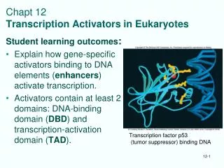
Regulation of transcription factor activity • The activity of transcription factors can be regulated by: (1) covalent modification (phosphorylation, acetylation, ubiquitination,) (2) by binding to ligands (nuclear receptors)
Phosphorylation of transcription factors Addition or removal of one or several phosphate groups on serine, threonine or tyrosine residues by a protein kinase or protein phosphatase.
Protein p53 • p53 (protein 53 kDa) is one of the most frequently cited biomolecules in Science Citation index • One of p53 functions is to act as a tumor supressor, which prevents cell division under DNA damaging conditions as exposure to UV light, etc • Knockout mice lacking p53 show normal development, but show predesposition to develop multiple tumors • About half of all 6.5 million people, annually diagnosed for various forms of cancer have mutations in p53 gene
Normal conditions DNA-damaging conditions protein kinase ATM p53 Phosphorylated p53 is stable and quickly accumulates p53 P P Unphosphorilated p53 not stable, p53 controlled genes not activated transcription transcription p53 P P DNA repair Apoptosis Arrest in G1 phase
Most p53 cancer associated mutaions occur in DNA binding regions