Download
1 / 70
720 likes | 869 Vues
Explore how transcription factors control gene expression in eukaryotes through DNA-binding, activation, and repression, with examples like SP1 & hormonal regulation. Learn about domain structures and regulatory elements influencing gene transcription.

E N D
Molecular Biology Course Section N Regulation of transcription in eukaryotes
N1 Eukaryotic Transcription Factors 1. Transcription factor domain structure: (DNA-binding, dimerization, transcription activation, repressor) 2. Targets for transcriptional regulation N2 Examples of transcriptional regulation SP1, hormonal regulation, phosphorylation of STAT proteins, HIV Tat, myoD homeodomain proteins
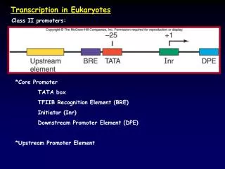
Section N: Regulation of transcription in eukaryotes N1 Eukaryotic Transcription Factors • Transcription factor domain structure (link) • DNA-binding domains • Dimerization domains • Transcription activation domains • Repressor domains • Targets for transcriptional regulation(link)
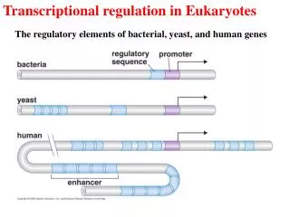
Transcription of a single gene may be regulated by many different factors interacting with regulatory elements upstream or downstream of the transcribed sequence. Start site Gene X +1 Regulatory elements to bind transcription factors
Example: The metallothionein (MT, 金属硫蛋白) gene The metallothionein protein protects the cell against excess concentrations of heavy metals, by binding the metal and removing it from the cell. The gene is expressed at a basal level, but is induced to greater levels of expression by heavy metal ions (such as cadmium) or by glucocorticoids (糖皮质素).
Common features of transcription factors • bind specifically to some DNA sites: specific motifs in promoters, upstream regulatory elements (UREs) or enhancer regions. Some factors modulate transcription by protein-protein intracation • Activate/repress transcription.
Transcription factors – domain structure Transcription factor Pdr1
- The activity of a transcription factor can be assigned to separate protein domains • activation domains. (activity) • DNA-binding domains. (activity) • dimerization domains. Many transcription factors occur as homo- or heterodimers, held together by dimerization domains. (regulation) • ligand-binding domains. Allowing regulation of transcription factor activity by binding of an accessory small molecule.The steroid hormone receptors are an example containing all for of these types of domain. (regulation)
Domain swap experiments moving domains among proteins, proving that domains can be dissected into separate parts of the proteins. The experiment of fusing activation domains of yeast transcription factors Gal4 and Gcn4 into the bacterial LexA repressor is described in your text book. Transcription activation domains are separable from their DNA binding activity.
Another example: construction of new proteins capable of binding to DNA NLS: nucleus localization signal
N1-2: DNA-binding domains • The helix-turn-helix domain • The zinc finger domain • The basic domain Return to menu…
Examples of Helix-turn-helix domains 1. Homeodomain: encoded by a sequence called the homeobox, containing a 60-amino-acid. In the Antennapedia transcription factor of Drosophila, this domain consists of four α-helices in which helices Ⅱand Ⅲ are at right angles to each other and are separated by a characteristic β-turn.
2. Bacteriophage DNA-binding proteins such as the phage λ cro repressor, lac and trp repressors, and cAMP receptor protein, CRP. • The recognition helix of the domain structure lies partly in the major groove and interacts with the DNA. • The recognition helices of two homeodomain factors Bicoid and Antennapedia can be exchanged, and this swaps their DNA-binding specificities.
Zinc finger domain exists in two forms. • C2H2 zinc finger: a loop of 12 amino acids anchored by two cysteine and two histidine residues that tetrahedrally co-ordinate a zinc ion. This motif folds into a compact structure comprising two β-strands and one α-helix. The α-helix containing conserved basic amino acids binds in the major groove of DNA (picture…picture2)
Examples: (1) TFIIIA, the RNA Pol III transcription factor, with C2H2 zinc finger repeated 9 times. (2) SP1, with 3 copies of C2H2 zinc finger. Usually, three or more C2H2 zinc fingers are required for DNA binding.
2.C4 zinc finger: zinc ion is coordinated by 4 cysteine residues. Example:steriod hormone receptor transcription factors (N2) consisting of homo- or hetero-dimers, in which each monomer contains two C4 zinc finger.(picture…)
The basic domain • Rich in basic amino acid residues • found in a number of DNA-binding proteins • generally associated with one or other of two dimerization domains, the leucine zipper or the helix-loop-helix(HLH) motif, resulting in basic leucine zipper (bZIP) or basic HLH proteins. Dimerization of the proteins brings together two basic domains which can then interact with DNA.
N1-3: Dimerization domains • Leucine zippers • The helix-loop-helix domain(HLH)
Leucine zippers • Leucine zipper proteins contain a hydrophobic leucine residue at every seventh position in a region that is often at the C-terminal part of the DNA-binding domain (picture.). • These leucines are responsible for dimerization through interaction between the hydrophobic faces of the α-helices. This interaction forms a coiled-coil structure
bZIP (basic leucine zipper) transcription factors: contain a basic DNA-binding domain N-terminal to the leucine zipper. The N-terminal basic domains of each helix form a symmetrical structure in which each basic domains lies along the DNA in opposite direction, interacting with a symmetrical DNA recognition site with the zippered protein clamp (pic1..) • The leucine zipper is also used as a dimerization domain in proteins containing DNA-binding domains other than the basic domain, including some homeodomain proteins.
The helix-loop-helix domain(HLH) • The overall structure is similar to the leucine zipper, except that a nonhelical loop of polypeptide chain separates two α-helices in each monomeric protein. • Hydrophobic residues on one side of the C-terminal α-helix allow dimerization. • Example: MyoD (pic..) family of proteins.
Similar to leucine zipper, the HLH motif is often found adjacent to a basic domain that requires dimerization for DNA binding. • Basic HLH proteins and bZIP proteins can form heterodimers allowing much greater diversity and complexity in the transcription factor repertoire.
N1-4: Transcription activation domains • Acidic activation domains • Glutamine-rich domains • Proline-rich domains
Acidic activation domains • Also called “acid blobs” or “negative noodles” • Rich in acidic amino acids • Exists in many transciption activation domains • yeast Gcn4 and Gal4, • mammalian glucocorticoidreceptor • herpes virus activator VP16 domains.
Glutamine-rich domains • Rich in glutamine • the proportion of glutamine residued seems to be more important than overall structure. • Exists in the general transcription factor SP1.
Proline-rich domains • Proline-rich • continuous run of proline residues can activate transcription • Exists in transcription factors c-jun, AP2 and Oct-2.
N1-5: Repressor domains • Repression of transcription may occur by indirect interference with the function of an activator. This may occur by: • Blocking the activator DNA-binding site (as with prokaryotic repressors,wrong) • Formation of a non-DNA-binding complex (e.g. the Id protein which blocks HLH protein-DNA interactions, since it lacks a DNA-binding domain, N2).
3. Masking of the activation domain without preventing DNA binding (e.g. Gal80 masks the activation domain of the yeast transcription factor Gal4). • A specific domain of the repressor is directly responsible for inhibition of transcription. (e.g. prokaryotic repressors) • e.g. A domain of the mammalian thyroid hormone receptor can repress transcription … (page 212 & 214). Return to menu…
N1-6: Targets for transcriptional regulation (pic…) chromatin structure; interaction with TFIID through specific TAFIIS; interaction with TFIIB; interaction or modulation of the TFIIH complex activity leading to differential posphorylation of the CTD of RNA Pol II.
It seems likely that different activation domains may have different targets, and almost any component or stage ininitiationand transcription elongation could be a target for regulation resulting in multistage regulation of transcription.Return to menu…
Section N: Regulation of transcription in eukaryotes N2 Examples of transcriptional regulation
Constitutive transcription factors:SP1 • Hormonal regulation:steroid hormone receptors • Regulation by phosphorylation:STAT proteins • Transcription elongation:HIV Tat • Cell determination:myoD • Embryonic development:homeodomain proteins
N2-1: Constitutive transcription factors:SP1 • binds to a GC-rich sequence with the consensus sequence GGGCGG. • binding site is in the promoter of many housekeeping genes • It is a constitutive transcription factor present in all cell types. • contains three zinc finger motifs and two glutamine-rich activation domains interacting with TAFII110, thus regulating the basal transcription complex.
N2-2: Hormonal regulation:steroid hormone receptors • Many transcription factors are activated by hormones which are secreted by one cell type and transmit a signal to a different cell type. • steroid hormones: lipid soluble and can diffuse through cell membranes to interact with transcription factors called steroid hormone receptors.
In the absence of steroid hormone, the receptor is bound to an inhibitor, and located in the cytoplasm (picture). • In the presence of steroid hormone, • the hormone binds to the receptor and releases the receptor from the inhibitor, • receptor dimerization and translocation to the nucleus. • receptor interaction its specific DNA-binding sequence (response element) via its DNA-binding domain, activating the target gene.
Steroid hormones involving important hormone receptors: glucocorticoid (糖皮质激素), estrogen (雌激素), retinoic acid (视黄酸)and thyroid hormone(甲状腺激素)receptors. Please noted that the above model is not true for all these hormone receptors • Thyroid hormone receptoris a DNA-bound repressor in the absence of hormone, which converted to a transcriptional activator.
N2-3: Regulation by phosphorylation: STAT proteins • For hormones that do not diffuse into the cell. • The hormones binds to cell-surface receptors and pass a signal to proteins within the cell through signal transduction. • Signal transduction often involves protein phosphorylation. Example: Interferon-γ induces phosphorylation of a transcription factor called STAT1α through activation of the intracellular kinase called Janus activated kinase(JAK). go on...
Unphosphorylated STAT1α protein: exists as a monomer in the cell cytoplasm and has no transcriptional activity. • Phosphorylated STAT1α at a specific tyrosine residue forms a homodimer which moves into the nucleus to activate the expression of target genes whose promoter regions contain a consensus DNA-binding motif (picture…pic3..)
N2-4: Transcription elongation:HIV Tat • Human immunodeficiency virus (HIV)(pic…) encodes an activator protein called Tat, which is required for productive HIV gene expression(pic..). • Tat binds to an RNA stem-loop structure called TAR, which is present in the 5’-UTR of all HIV RNAs just after the HIV transcription start site, to regulate the level of transcription elongation.
In the absence of Tat, the HIV transcripts terminate prematurely due to poor processivity of the RNA Pol Ⅱ transcription complex. • Tat binds to TAR on one transcript in a complex together with cellular RNA-binding factors. This protein-RNA complex may loop backwards and interact with the new transcription initiation complex which is assembled at the promoter. go on...
This interaction may result in the activation of the kinase activity of TFIIH, leading to phosphorylation of the carboxyl-terminal domain (CTD) of RNA PolⅡ, making the polymerase a processive enzyme to read through the HIV transcription unit, leading to the productive synthesis of HIV proteins (picture..)
N2-5: Cell determination:myoD(pic1..pic2..) • myoD was identified as a gene to regulate gene expression in cell determination, commanding cells to form muscle. • MyoD protein has been shown to activate muscle-specific gene expression directly. Overexpression of myoD can turn fibroblasts into muscle-like cells which express muscle-specific genes and resemble myotomes. • myoD also activates expression of p21waf1/cip1 expression, a small molecule inhibitor of CDKs, causing cells arrested at the G1-phase of the cell cycle which is characteristic of differentiated cells. .
Four genes,myoD,myogenin, myf5 and mrf4 have been shown to have the ability to convert fibroblasts into muscle. The encoded proteins are all members of the helix-loop-helix (HLH for dimerization) transcription factor family. • These proteins are regulated by an inhibitor called Id that lacks a DNA-binding domain, but contains the HLH dimerization domain. Id protein can bind to MyoD and related proteins, but the resulting heterodimers cannot bind DNA, and hence cannot regulate transcription
N2-6: Embryonic development: homeodomain proteins • The homeobox is a conserved DNA sequence which encodes the helix-turn-helix DNA binding protein structure called the homeodomain. • Homeotic genes of Drosophila are responsible for the correct specification of body parts. For example, mutation of one of these genes, Antennapedia, causes the fly to form a leg where the antenna should be. • conserved between a wide range of eukaryotes. • important in mammalian development.