Download
1 / 52
750 likes | 3.4k Vues
Jaundice and its Investigation. Andrew M Smith Jan 2011. Jaundice. "it looks like there's something wrong… ….with your television set.“ Matt Groenig, creator of The Simpsons. Jaundice. An elevation of serum bilirubin above normal limit (9 mmol/l)

E N D
Jaundice and its Investigation Andrew M Smith Jan 2011
Jaundice "it looks like there's something wrong… ….with your television set.“ Matt Groenig, creator of The Simpsons
Jaundice • An elevation of serum bilirubin above normal limit (9 mmol/l) • Clinically evident at ~ 35 mmol/l
Objectives • Review Liver Anatomy and Physiology • Classification and causes of Jaundice • Investigation of Jaundice • Principles of Management of Jaundice • Cases • Summary
Functions of the Liver • 1.Metabolism • Fats • Proteins • Carbohydrates • Hormones • 2.Storage • 3.Metabolism and excretion bilirubin • 4. Drug metabolism and excretion
Normal Bile Physiology • 250-500 mg bile/day • Water (98%) • Bile Salts • Bile pigments (Bilirubin) • Fatty Acids • Lecithin • Cholesterol
Normal Bilirubin Metabolism RBC Hb Degraded to Globin + Fe + Bilirubin Hepatocyte Conjugated Bilirubin Diglucuronide Bilirubin bound to albumin Kidney Urobilinogen Portal Vein Urobilinogen Intestine Bilirubin Urobilinogen Stercobilin
Major Causes of Jaundice Pre-hepaticHaemolysis Ineffective erythropoiesis HepaticPrematurity Gilberts Drugs Hepatitis: viral, NASH Alcohol / cirrhosis Tumours Extrahepatic sepsis Post-hepatic ‘Obstructive’Gallstones (in the lumen) Bile duct stricture ( in the wall) Ca pancreas (extrinsic)
Investigation Of A Jaundiced Patient • History • Examination • Tests • Blood • Urine • Imaging
History ‘most important part of the evaluation of the patient with jaundice’
History 1. Jaundice – onset 2. Pale stools, dark urine? YES = POST HEPATIC NO = PRE & HEPATIC PAIN? YES NO Pre-hepatic: Family history of bleeding disorders, tendency to bleed Wt loss Back Pain Non-specific symptoms MALIGNANCY Colicky Fatty food intolerant GALLSTONES Hepatic: IV Drug abuse blood transfusions Travel flu-like illness Excess alcohol intake Obesity Drug History Hepatitis ASSOCIATED FEVERS / RIGORS? Gram –ve Septicaemia ADMIT Cirrhosis/ NASH
Examination • Stigmata Chronic Liver disease • Hepatomegaly – texture,edge, nodules • Hepatosplenomegaly • Ascites –shifting dullness • Portal hypertesion • Obvious iv drug use
Examination – obstructive jaundice • Temp • Tachycardic +/- hypotensive • Cachexia, Virchow’s node,clubbing • Murphy’s sign • Courvoisier’s law ‘If in the presence of jaundice the gallbladder is palpable then the cause of the jaundice is unlikely to be gallstones’ • Urine cholangitis
Investigations for jaundice • Bloods • General – Liver Function Tests - Albumin, INR (give more info on function!) • Specific • Urine • Imaging • Histology
Ix Jaundice – Bloods • Liver Function Tests - really a test of hepatocyte damage Alanine Transaminase ALT range <40iu/L elevated cellular damage AlkalinePhosphatase ALP range 70-300iu/KL elevation post hepatic obstruction Bilirubin range 5- 40 umol/L
Prehepatic • Unconguated Bil ↑ • LFT’s N • haptoglobins ↓ • Reticulocytes ↑ • Coombs test +ve • Clotting screen • Urine urobilinogen↑
Hepatic • ALT ↑ ↑ ↑ • ALP N or ↑ • Bil ↑ • Albumin ↓ • INR ↑ • Hepatitis serology • Autoantibodies • Anti-mitochondrial PBC • Anti-nuclear & antimicrosomal, Autoimmune hepatitis • Caeruloplasmin ↑ • Wilson’s • γ-Globulins ↑ • Cirrhosis esp autoimmune • Transferrin ↑↑ • Haemochromatosis ↑ • α-foetoprotein, αFP ↑ • HCC in cirrhosis
Post - hepatic • ALT N or ↑ • ALP ↑ ↑ ↑ • Bil ↑ • INR ↑ • CEA, Ca19.9 ↑ • Panc & cholangio Ca
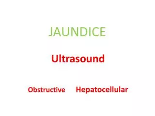
Imaging - Ultrasound • Key investigation • Distinguish hepatic and post hepatic • Identify gallstones
Imaging - Ultrasound Key information from report BILIARY DUCT DILATION Calculi Gallstones present, GB wall thickness CBD diameter normal (<7mm) No calculi No gallstones, but CBD ↑ ? Pancreatic malignancy NO DUCT DILATION Texture of liver eg normal, fatty, micronodular Lesions present
Imaging - Endoscopic ultrasound CBD CBD PD PD
Investigation Summary • First line • LFT’s & USS • Second line • If dilated ducts refer for stone or ? maligancy management • No ducts – parenchymal liver disease • Ensure good alcohol history • Hepatitis serology • Hepatic autoantibodies • Ferritin
Case 1 • A 18 year old student comes to see you and reveals that his mates taunt him as he often appears to have yellow eyes? • What do you do?
Gilbert’s disease • Diagnosis of exclusion • Good Hx. No family hx of sickle/G6PD defficiency • no other risk factors • Notes jaundice worsens on fasting • Unconguated Bil ↑ and LFT’s N • haptoglobins Reticulocytes both normal, Coombs test -ve 5 -7 % population, reasssure.
Case 2 • A Samuel Smiths delivery man who enjoys the companys perks to excess attends, complaining of a distended abdomen which is becoming painful? • Diagnosis? • Management?
Decompensated alcoholic cirrhosis History – confirms 100+ unit intake for 20 yrs Examination – stigmata chronic liver disease abdo, palpable liver and spleen shifting dullness Ix - LFTs Bil ↑ , ALT ↑ ↑ ↑, ALP ↑ INR ↑ Albumin low USS , cirrhosis, splenomegaly and ascites Treatment – Cessation of alcohol - treatment of withdrawal - thiamine, folic acid - low salt diet, spironolactone - Liver bx when ascites settles - Ix portal htn, OGD, banding, B –blocker, TIPs
Case 3 • You are asked to make a home visit to see a 53 yr old man with severe abdominal pain . His notes show that he had an episode of pancreatitis on holiday in Spain a year ago. • He tells you that the has had upper tummy pain, can’t get comfortable and has had shakes and feels cold? • What is the diagnosis? • What action do you take?
Ascending Cholangitis • Examination reveals fever, jaundice and a tachycardia. • He has Charcot’s triad – pain, jaundice, fever, ie ascending cholangitis • He needs an emergency admission, significant morbidity and mortality • iv access, analgesia
Ascending Cholangitis Athospital, continue resuscitation, antibiotics, check and correct INR Emergency ERCP and duct clearance Laparoscopic Cholecystectomy, same admission
Gallstones • Previous pancreatitis due to gallstones. 20% incidence of further complications within 6 months once symptomatic • In elective situation can avoid ERCP, by performing a duct exploration at the time of laparoscopic cholecystectomy • On horizon of further sea change with advent of NOTES (natural orifice transluminal endoscopic surgery)
Case 4 • A 37 year old Chinese immigrant who has just arrived in Leeds, presents frankly jaundiced with a history of abdominal pain and weight loss. On examination he is clearly jaundiced and has a palpable liver. • What do we do next? • Can we make an educated guess from the history?
Hep C and HCC • LFT’s and USS – ALT, ALP and Bilirubin grossly elevated. • USS cirrhosis and multiple lesions. Referred. • CT and Hep C, aFP Beyond transplant or resection Rx Chemoembolisation / BSC
Case 5 Your senior partner has been seeing for a year a previously fit 43yr old man with non specific symptoms of fatigue. Two consecutive ALT’s six months apart were elevated at 120, and 107 ( normal < 40). The rest of his blood work was normal. Do you act on this result?
Investigation isolated raised ALT • Present > 6 months should investigate • Good Hx and Exam FIRST WAVE TESTS 1 .Exclude drugs NSAIDs, antibiotics, statins, antiepileptic drugs anti-TB. Herbal remedies. Paracetamol 2. Assess Alcohol excess 3. Hep B and C 4. Hereditary Haemochromotosis 5. NASH and steatosis SECOND WAVE TESTS Refer 6. Thyroid/Coeliac/muscle disorders THIRD WAVE – Definitely refer
What is the most likely cause of jaundice that I will see? South Wales, Gut 2002 Glasgow, Gut, 2002 Alcoholic liver disease Gallstones Malignacy
Summary • Good history will direct rest of care • LFTs and USS initially • Admit cholangitis when suspected • Admit for symptom control
Hep B • Send hepatitis serlogy . • Will assess status to determine whether immune/carrier or chronic infection • HepBsAg, HepBsAb, HepBcAb • chronic infection HepBsAG +ve + HepBcAb +ve • immune HepBsAb +ve , HepBcAb +ve • HBV DNA
Hep C • Hep C Antibody • Then Hep C RNA, Hep C genotype and liver biopsy
Haemochromotosis • Frequency 5/1000 • Fe and TIBC, • Fe saturation > 45% then ferritin • Ferritin > 400ng/ml • Liver biopsy
NASH • NASH more common women and type 2 Diabetes • Hep B/C/HCC negative USS to look for steatosis • Bx if stigmata chronic liver disese
Isolated Hyperbilirubinaemia • Occurs – excess production or impaired uptake • Check conjugated vs unconjugated • Assess Haemolysis • No haemolysis, fluctuating bilirubin – gilberts disease.